Lophiomurispora hongheensis Wanas. sp. nov. (Figures 7 and 8)
MycoBank number: MB 837998; Index Fungorum number: IF 837998; Facesoffungi number: FoF 11582;
Etymology: The specific epithet is derived from Honghe County, the region of Yunnan Province in which this species was gathered.
Holotype: HKAS110127
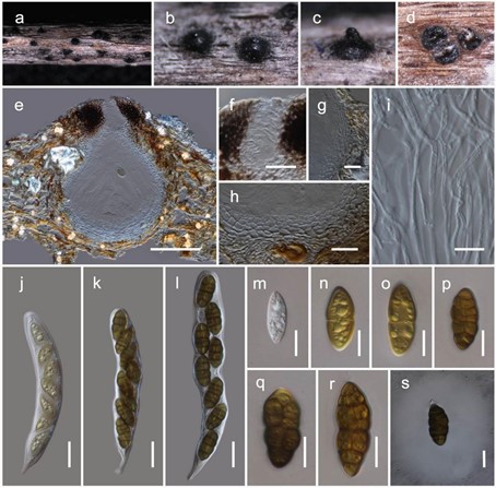
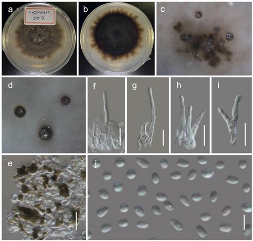
It is saprobic on dead twigs of Dodonaea viscosa Jacq. (Sapindaceae) in terrestrial habitats. Sexual morph: The ascomata is a 280–360 µm high, 200–250 µm diam. (M = 318.6 × 232.7 µm, n = 5), scattered to gregarious, immersed, coriaceous, dark brown to black, globose to subglobose ostiolate. The ostiole is a 70–100 µm long, 40–80 µm diam. (M = 82.1 × 64.8 µm, n = 5), crest-like, central papillate, with a pore-like opening, comprising hyaline periphyses. The peridium is 20–30 µm wide at the base, 30–60 µm wide at the sides, broad at the apex, comprising two strata, with outer stratum composed of small, pale brown to brown, slightly flattened, thick-walled cells of textura angularis, fusing and indistinguishable from the host tissues. The inner stratum is composed of several layers with lightly pigmented to hyaline cells of textura angularis to textura prismatica. The hamathecium comprises 1–2 µm wide, branched, septate, cellular pseudoparaphyses, situated between and above the asci, embedded in a gelatinous matrix. The asci are 120–160 × 17–22 µm (M = 135.2 × 18.5 µm, n = 15), eight-spored, bitunicate, fissitunicate, cylindric-clavate, with a short pedicel, and is rounded at the apex, with an ocular chamber. The ascospores are 25–30 × 11–13 µm (M = 27.8 × 12 µm, n = 30), uni- to bi-seriate, overlapping, and are initially hyaline, turning brown at maturity. They are ellipsoidal to fusiform, muriform, four-to-eight-transversely septate, with one-to-two-longitudinal septa. They are slightly curved, deeply constricted at the central septum, slightly constricted at the remaining septa, conically rounded at the ends, and smooth-walled, with a distinct mucilaginous sheath. Asexual morph: Coelomycetous. The conidiomata is 1–1.5 mm diam. pycnidial, phoma-like, solitary, gregarious, dark brown to black, and immersed, with a sphaerical mass of slimy conidia oozing out at ostiolar apex. The conidiomata wall is multi-layered, with brown-walled pseudoparenchymatous cells, with a hyaline inner most layer. The conidiophores are 10–15 × 1.5–2.5 µm long (M = 12.4 × 2.1 µm, n = 15), septate and sparsely branched, which are formed from the inner most layer of the pycnidium wall. The conidiogenous cells are phialidic, cylindrical, hyaline, flexuous and smooth, with a short collarette. The conidia are 2.5–4 × 1.5–2 µm (M = 3 × 1.7 µm, n = 50), hyaline, aseptate, straight to curved, ellipsoidal with rounded ends, and are thin-walled, smooth-walled, and numerous
Culture characteristics: the colonies on PDA reached a 4 cm diameter after 2 weeks at 20 ◦C. They were circular, had a serrate margin, and were whitish at the beginning, becoming greenish-brown 4 weeks later. They were slightly raised, and reverse dark brown. The hyphae septate were branched, hyaline, thin, and smooth-walled.
Known distribution: Yunnan, China, on Dodonaea viscosa.
Material examined: China, Yunnan, Honghe Hani and Yi Autonomous Prefecture, Honghe County, 23.421068 N, 102.229128 E, 735 m, on dead twigs of Dodonaea viscosa, 22 April 2020, D.N. Wanasinghe, Honghe 003 (HKAS110127, holotype), ex-type culture, KUMCC 20-0217, ibid. 23.419206 N, 102.231375 E, 618 m, Honghe 008 (HKAS110129, paratype), ex-paratype living culture, KUMCC 20-0223, ibid. 23 April 2020, ibid. DWHH07-1 (HKAS110130), living culture, KUMCC 20-0224, DWHH01 (HKAS110132), living culture, KUMCC 20-0216, ibid. DWHH04 3 (HKAS110131), living culture, KUMCC 20-0219. Parapyrenochaetaceae Valenz-Lopez, Crous, Stchigel, Guarro and J.F. Cano, Studies in Mycology 90: 64 (2017)

Figure 7. Sexual morph of Lophiomurispora hongheensis (HKAS110127, holotype). (a–c) Ascomata on the dead woody twigs of Dodonaea viscosa; (d) cross section of ascomata; (e) vertical section of ascoma; (f) closeup of ostiole; (g,h) peridium; (i) pseudoparaphyses; (j–l) asci; (m–s) ascospores (s in Indian Ink); Scale bars, (e) 100 µm; (f–h,j–l) 20 µm; (i,m–s) 10 µm.

Figure 8. Asexual morph of Lophiomurispora hongheensis (KUMCC 20-0217, ex-type culture). (a,b) colony on PDA (b from the bottom); (c,d) immersed pycnidia in PDA (from the bottom); (e) pycnidia wall; (f–i) conidiophore; (j) conidia. Scale bars, (e–i) 10 µm; (j) 5 µm.
