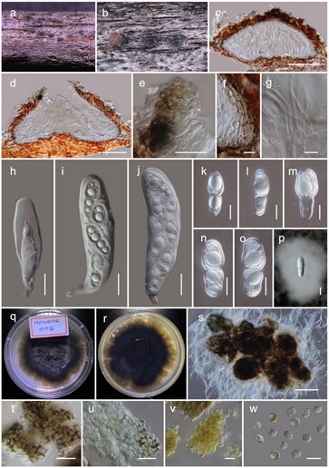
Haniomyces dodonaeae Wanas. & Mortimer, in Wanasinghe, Mortimer & Xu, Journal of Fungi 7(3, no. 180): 25 (2021) (Figure 5)
MycoBank number: MB 837997; Index Fungorum number: IF 837997; Facesoffungi number: FoF 11379;
Etymology: The specific epithet reflects the host genus Dodonaea.
Holotype: HKAS110128
It is saprobic on dead twigs of Dodonaea viscosa Jacq. (Sapindaceae). Sexual morph: the ascomata is a 150–200 µm high, 350–450 µm diam. (M = 165.4 × 390.3 µm, n = 5), scattered, semi-immersed to erumpent, subglobose to conical or shaped irregularly, flattened base, glabrous, brown to dark brown ostiolate, fused with host tissues. The ostiole is a short papillate, black and smooth, with hyaline periphyses (15–25 µm long, 1.5–2 µm wide). The peridium 5–10 µm wide at the base, 10–20 µm wide at sides, comprising 2–4 layers, outer layer pigmented, comprising reddish-brown to dark brown, with thin-walled cells of textura angularis, and an inner layer composed of hyaline, loosen, cells of textura angularis. The hamathecium comprises numerous, 2–3 µm wide, filamentous, branched, septate, pseudoparaphyses. The asci are 110–130 × 25–35 µm (M = 118.5 × 31.2 µm, n = 20), eight-spored, bitunicate, fissitunicate, clavate, with a short pedicel (10–15 µm long), apically rounded with an ocular chamber. The ascospores 25–35 × 12–15 µm (M = 32.2 × 14.3 µm, n = 30), overlap the biseriate, are ellipsoidal to sub-fusiform, hyaline, one-septate, with the septum almost median, deeply constricted at the middle septum, with the upper cell wider than the lower cell, and are smooth-walled with small to large guttules in each cell, rounded at both ends and covered by a distinct mucilaginous sheath (30–50 µm, diam.). Asexual morph: Coelomycetous. The conidiomata are up to 250 µm diam., sporodochial on PDA, globose, solitary or aggregated, semi-immersed, black, exuding yellow conidial masses. Conidiophores and conidiogenous cells were not observed in vitro. The conidia are 5.5–7.5 × 4.5–5.5 µm (M = 6.4 × 5.4 µm, n = 30), solitary, aseptate, globose or ellipsoid, with the hyaline becoming medium to golden brown and finely verruculose.
Culture characteristics: the colonies on PDA reached a 3 cm diameter after 2 weeks at 20 ◦C. They were circular has a serrate margin, whitish at the beginning, becoming brown at the centre and brownish-green towards the margin after 4 weeks. They were slightly raised, and reverse blackish brown. The hyphae septate were branched, hyaline, thin, and smooth-walled.
Known distribution: Yunnan, China, on Dodonaea viscosa.
Material examined: China, Yunnan, Honghe Hani and Yi Autonomous Prefecture, Honghe County, 23.421068 N, 102.229128 E, 735 m, on dead twigs of Dodonaea viscosa, 22 April 2020, D.N. Wanasinghe, Honghe 005 (HKAS110128, holotype), ex-type living culture, KUMCC 20-0220, ibid. 23.419206 N, 102.231375 E, 618 m, Honghe 010 (HKAS110125, paratype), ex-paratype living culture, KUMCC 20-0221.

Figure 5. The sexual (HKAS110128, holotype) and asexual (KUMCC 20-0220, ex-type) morphs of Haniomyces dodonaeae. (a,b) ascomata on the dead woody twigs of Dodonaea viscosa; (c,d) vertical section of ascoma; (e) periphyses; (f) peridium; (g) pseudoparaphyses; (h–j) asci; (k–p) ascospores (p in Indian Ink); (q,r) colony on potato dextrose agar (PDA) (r from the bottom); (s) squashed pycnidia which were produced on PDA; (t) pycnidia wall; (u–w) conidia. Scale bars, (c,d) 100 µm; (e,h–j,t,u) 20 µm; (f,k–p,v,w) 10 µm; (s) 200 µm.
