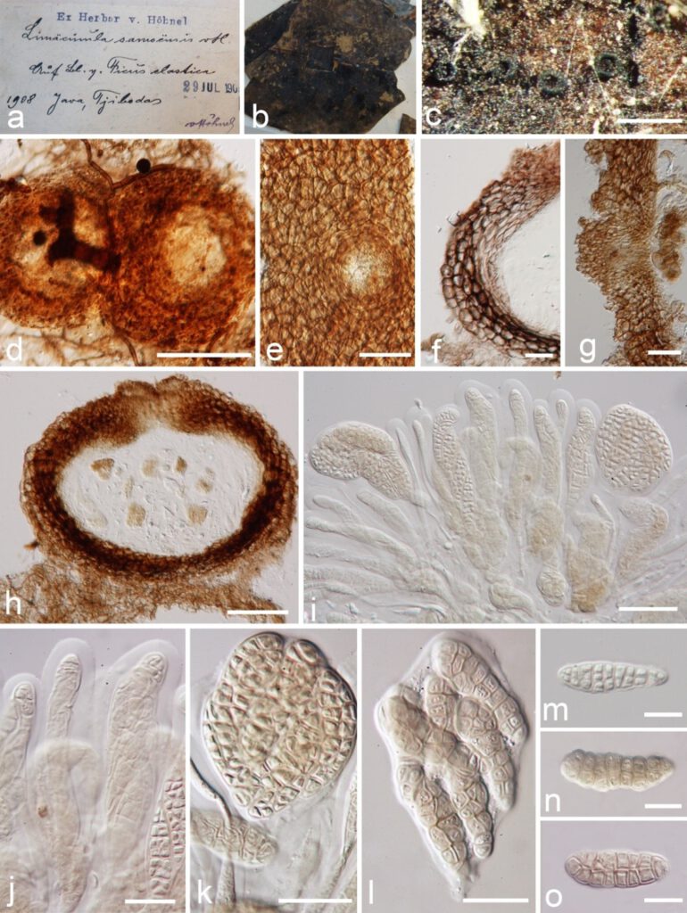
Limacinula samoensis Höhn. [as ‘samoënsis’], Sber. Akad. Wiss. Wien, Math.naturw. Kl., Abt. 1 118: 1200 (1909), Figure 19
MycoBank number: MB 158863; Index Fungorum number: IF 627637; Facesoffungi number: FoF 10357;
Saprobic, Epiphytic or biotrophic on the leaves of Ficus elastica, mixed with other fungal taxa, as sooty molds adpressed to the surface of host gaining nutrients from sugary exudates of sap-feeding insects. Subiculum well-developed, superficial, loose, brown, comprising effuse, branched, subhyaline to brown, septate hyphae, reticulate. Sexual morph: Ascomata 135–220 μm in diam. (x̅ = 155 μm, n = 10), superficial, sessile on subiculum, perithecial, scattered to aggregate, globose, collabent when mature, uniloculate, brown to black, with periphysate ostiole, thick-walled, more or less setose. Wall of ascoma 32–48 μm (x̅ = 40 μm, n = 10), multi-layered, externally comprising pigmented, dark brown, thick-walled cells of textura angularis, with inner layer thinner, composed of irregularly-shaped, flattened, lightly brown to hyaline, thin-walled cells of textura angularis. Hamathecium lacking paraphyses. Asci 58–115 × 22–52 μm (x̅ = 87 × 38 μm, n = 10), 8-spored, bitunicate, fissitunicate, clavate when immature, obpyriform to obovoid at maturity, shortly pedicellate or sessile, lacking a distinct ocular chamber. Ascospores 22–35 × 8–12 μm (x̅ = 30 × 9.5 μm, n = 10), overlapping uni-seriate to multi-seriate, irregularly arranged, fusoid to oblong, basal cells thinner than upper cells, rounded at both ends, hyaline to light brown, muriform, with 6–8 transverse septa, 3–6 longitudinal septa, constricted at the septum, slightly constricted at the septum, smooth and thick-walled, lacking a gelatinous sheath or appendages. Asexual morph: Undetermined.
Material examined: Indonesia, Java, on leaves of Ficus elastica Roxb. ex Hornem. (Moraceae), 1908, von Höhnel (K, Ex Herbarium von Höhnel).

Figure 19 Limacinula samoensis (K, Ex Herbarium von Höhnel). a, b Herbarium material. c Ascomata on the superficial of the host. d, e Squash mounts of ascoma. f Vertical section through ascoma wall. g Vertical section through ostiole. h Vertical section of ascoma. i–l Asci with ascospores. m–o Ascospores. Scale bars: c = 500 µm, d =100 µm, e, h = 50 µm, f, g, i = 25 µm, j–l = 20 µm, m–o = 10 µm.
