Ligninspaheria jonesii J.F. Zhang, J.K. Liu, K.D. Hyde & Z.Y. Liu, sp. nov., Index Fungorum number: IF551758
Etymology: Named in honour of E. B. Gareth Jones for his contributions to tropical mycology.
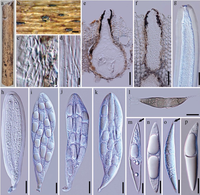
Saprobic on decaying bamboo culms, forming black spots on the host surface. Sexual morph: Ascomata 353–541 µm high (including neck), 298–317 µm diam, scattered, solitary, immersed under the host tissue, black, clypeate, subglobose to obpyriform, visible on host surface as raised, dark spots. Ostiole central, long, slit-like opening, lacking periphyses in ostiolar canal. Peridium up to 19–37 µm wide, coriaceous to carbonaceous, composed of two strata, an outer stratum of thick-walled and brown cells arranged in a textura angularis and fusing with host cells, and inner layer of hyaline cells of textura angularis. Hamathecium up to 3–6 µm wide, comprising dense, cellular pseudoparaphyses, anastomosing above and between the asci, embedded in a gelatinous matrix. Asci 163–243 × 38–57 µm (x̅ = 208 × 48 µm, n = 20), 8-spored, bitunicate, fissitunicate, broad-clavate with tapering pedicel, apically rounded, with a distinct ocular chamber. Ascospores 79–121 × 14–23 µm (x̅ = 97 × 18µm, n = 40), 3–4-seriate, hyaline, indistinctly 1-septate when mature, broadly fusiform, slightly curved in the center, pointed at both ends with a 3 µm long, 2.5 µm wide, gelatinous cap. Asexual morph: Undetermined.
Culture characters: Ascospores germinating on WA within 24 h and germ tubes produced from ends. Colonies growing fast on PDA, reaching 23 mm in 2 weeks at 25 °C, flat or raised at the center, circular, irregular at the margin, grayish from above, dark brown from below. Mycelium immersed in the media, composed of branched, septate, smooth, grayish hyphae.
Material examined: THAILAND, Chiang Rai, Muang District, on limestone outcrops, Mae Chang Hot Spring, on decaying bamboo culms, 25 Nov 2014, j. F. Zhang (holotype MFLU 15-2718, ex-type living culture, MFLUCC 15-0641); ibid., 25 Nov 2014, J.F Zhang (GZAAS 15-0115); living culture GZCC 15-0080.
Notes: Ligninspaheria is clearly different from other groups within the order of Pleosporales based on molecular data and morphological characters. This monotypic genus is introduced to accommodate those taxa characterized by having deeply immersed ascomata, surrounded by a large blackened clypeus, with long immersed ostioler canal and broad-clavate asci with fusiform ascospores surrounded by a gelatinous cap at both ends. Ligninsphaeria jonesii is similar to Pseudotricha guatopoensis in having immersed, obpyriform ascomata and clavate asci. However, the former is distinct in pseudoparaphyses type (cellular vs. trabeculate), ascospore appendages (gelatinous cap at both ends vs. 5–6 µm wide gelatinous sheath) and ascospore septation (1-septate vs. 3–5-septate) (Huhndorf et al. 1994). The new fungus is also similar with Lophiotremataceae, but differs by its broad-clavate ascus and ascospores surrounded by a gelatinous cap at both ends, while the ascus in the latter taxon is cylindrical and ascospores lack gelatinous appendages (Hirayama & Tanaka 2011). In addition, the molecular analysis showed that this new fungus is phyl==ogenetically close with Astrosphaeriella-like taxa, as well as the families Delistchiaceae and Testudinaceae. However, they have different morphological characters. Morphological characters of this new fungus are obviously different from other genera even families within the Pleosporales, but the phylogenetic placement of these groups is not stable, which may be caused by the lack of molecular data for taxa in these groups. Moreover, only one collection was obtained in this study, which cannot commendably illustrate the phylogenetic relationship with close groups. Therefore, we introduced a new genus Ligninsphaeria to accommodate the new fungus and future studies in tropical regions are likely to collect and provide data for this undersampled group. The recent study on Astrosphaeriellaceae and Pseudoastrosphaeriellaceae is a good example (Phookamsak et al. 2015).
Fig. 2 Ligninsphaeria jonesii (MFLU 15-2718) a The host, a decaying bamboo culm. b Ascomata immersed in the bamboo host. c
Section of peridium. d Pseudoparaphyses. e Vertical section through ascoma. f Long immersed ostiole. g Asci with fissitunicate dehiscence.
h–k Asci. l Germinating ascospore. m–p Ascospores. Note the arrowheads indicate the gelatinous cap on both ends in m and o. Scale bars:
b = 200 µm, c, d = 5 µm, e = 100 µm, f = 50 µm, g–h, n–p = 20 µm, i–k, m = 10 µm, l = 30 µm.