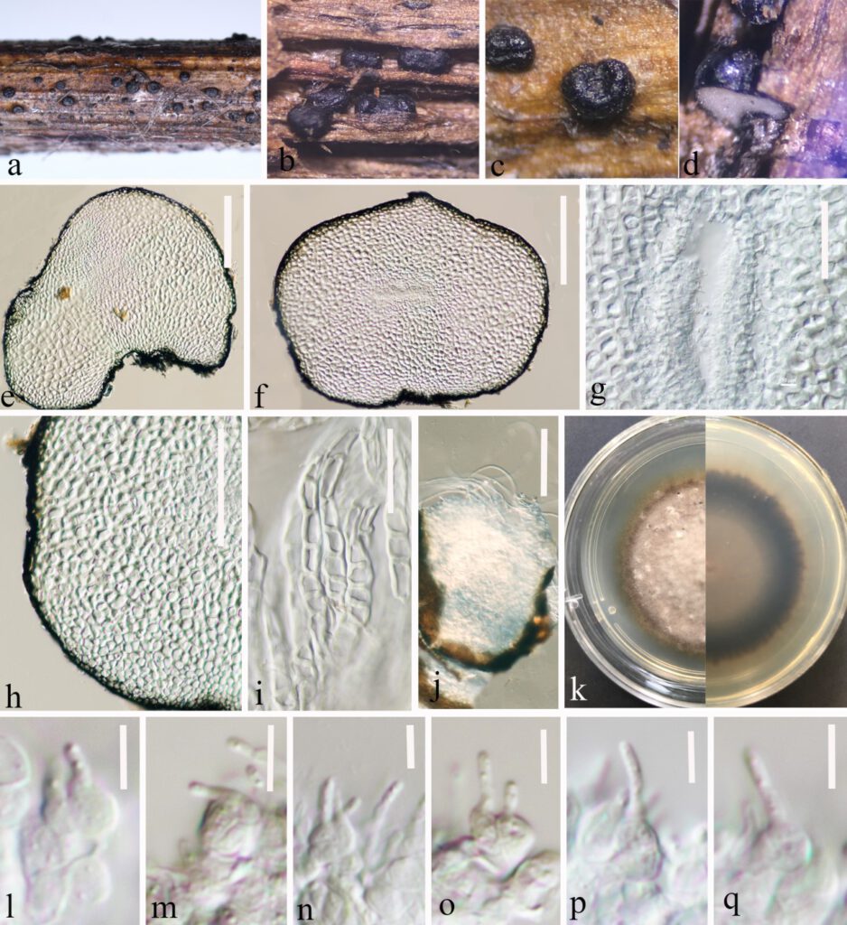
Leptosphaeria yunnanensis Y. Gao, H. Gui & K.D. Hyde, sp. nov.
MycoBank number: MB; Index Fungorum number: IF; Facesoffungi number: FoF 12905; FIGURE5. ZG25.
Etymology: The specific epithet “yunnanensis” refers to Yunnan Province where the holotype was collected.
Holotype: HKAS 124670
Saprobic on decaying stalk of grass. Sexual morph: Undetermined. Asexual morph: Conidiomata 280–515 μm diam × 170–370 μm high (x̅ = 385 × 253 μm, n = 20), solitary, scattered or in small groups, erumpent to superficial, globose to subglobose, small to medium-sized, smooth-walled, easily removed from the host substrate, black, coriaceous, without ostiolate. Conidiomatal wall (132–)144–189(–202) μm thick, (x̅ = 166 μm, n = 30), thickness, almost fills the entire conidiomata, each cell-layer (9–)10.6–15.6(–18) μm wide, (x̅ = 13 μm, n = 40), composed of flattened cells of textura angularis, heavily pigmented in the outer layers, lined with a hyaline innermost layer bearing conidiogenous cells. Conidiogenous cells (2.8–)3.6–5.2(–6.4) μm long × (3.8–)4.4–5.8(–6.7) μm high (x̅ = 4.4 × 5 μm, n = 40), ampulliform or globose to subglobose, smooth-walled, hyaline. Conidia (2.5–)3.3–5.6(–6.6)μm long × (1–)1.2–1.5(–1.8)μm wide (x̅ = 4.5 × 1.4μm, n = 30), ellipsoidal to sub-cylindrical with obtuse ends, hyaline, aseptate, smooth-walled.
Culture characteristics: conidiomata germinated on PDA within 20 hours. Colonies on PDA reaching 20 mm in 4 weeks at room temperature (25–27°C), hairy or cottony, raised, white to grey, mycelium superficial, dark brown at margin, white to light grey at centre from the above, grey in the center gradually black towards the edges from the below.
Material examined: CHINA, Yunnan Province, Zhaotong city, Daguan County, Grassland (27°44’23”N, 103°47’59”E), on decaying stalk of herbaceous plant, 21 August 2021, Ying Gao, ZG25A (HKAS 124670, holotype), ex-type living culture, CGMCC 3.23748. ZG25C (HKAS 124671, paratype), ZG25B ex-paratype living culture CGMCC3.23749
Note: Based on the multi-gene phylogenetic analyses, Leptosphaeria yunnanensis clusters between Leptosphaeria italica (MFLU15-0174), Leptosphaeria urticae (MFLU 18-0591) and Leptosphaeria pedicularis (CBS 390.80). The nucleotide pairwise comparison showed that Leptosphaeria yunnanensis differs from Leptosphaeria urticae in 44/487 bp of ITS (9.03%) and Leptosphaeria pedicularis (CBS 390.80) in 37/512 bp of ITS (7.23 %), 24/334 bp of tub2 (7.19 %). Following the guidelines for species delineation described by Jeewon and Hyde (2016) and Leptosphaeria yunnanensis differs from all previously known species of the genus in morphological description (Gruyter et al. 2013, Niranjan et al. 2018, Pem et al. 2020a, Lestari et al. 2021). Leptosphaeria yunnanensis is unique in conidiomatal wall which occupied almost the entire interior of the conidiomata, and sporulation indistinctly in the center. Therefore, we introduce Leptosphaeria yunnanensis as a novel taxon

FIGURE5 ZG25. Leptosphaeria yunnanensis (HKAS 124670 holotype). a–d. Black conidiomata on the host surface. e, f. Vertical sections of conidiomata. g. Conidia location in conidiomata. h–i. Peridium. j. A germinating conidiomata. k. Front and reverse of colony on PDA. l–q. Conidiogenous cells and developing conidia in conidiomata. Scale bars:e, f = 150 μm. g = 50 μm. h = 100 μm. i = 50 μm. j = 100 μm. l–q = 5 μm.
