Lepiota venenata Zhu L. Yang & Z.H. Chen, in Cai, Chen, He, Luo & Yang, J. Fungal Res. 16, 67 (2018)
IndexFungorum number: IF 824765; MycoBank number: MB 824765; Facesoffungi number: FoF 10409; Figs. 86, 87
Pileus 17–20 mm diam., first parabolic, expanding to umbonate with high umbo, with inflexed margin; fibrillose squamules and squamules at umbo towards the margin, light brown to brown (7D6–8), slightly darker at the umbo, on white fibrillose background; marginal zone with brown concolorous fibrillose squamules and white thin fibrillose. Lamellae free, crowded, ventricose, 2–3 mm wide, white, with concolourous eroded edge. Stipe 25–32 × 3–4 mm, cylindrical, with slightly tapering to apex; covered with light brown to brown (7D6–8) squamules from annular zone toward the base on white background. Annular zone, with white fibrillose and concolourous squamules. Context white in pileus, 1.5–2 mm wide; white and hollow in stipe. Smell and taste not observed. Spore print white.
Basidiospores [50,1,1] 5.5–7.5 × 3.5–4.5 µm, avl × avw = 6.5 × 4.0 µm, Q=1.5–1.8, Qav= 1.6, oblong ovoid in side view, oblong in frontal view, hyaline, slightly thick-walled, dextrinoid, congophilous, cyanophilous, not metachromatic in Cresyl Blue. Basidia 15–18× 5.0–6.5 µm, clavate, thin-walled, hyaline, 4-spored. Pleurocystidia absent. Cheilocystidia 15–25 × 5.0–8.0 µm, mostly clavate, often utriform, rarely cylindrical, hyaline, thin-walled. Pileus covering a trichoderm made up of cylindrical elements, always tapering to the apex, 55–275 × 6.0–13 µm, slightly thick-walled, with brown intracellular and parietal pigment, without short clavate or clavate elements under long elements, branched and septate. Clamp connections present in all tissues.
Material examined – Laos, Xekong Province, Tha Teng District, 30 September 2015, P. Sysouphanthong (HNL503248).
GenBank accession numbers – HNL503248 – ITS: MZ717724.
Known habitat – grow solitary to a small group, terrestrial on rich humus soil (Cai et al. 2018, this study).
Known distribution (based on molecular data) – China (Cai et al. 2018), central Laos (this study).
Notes – Lepiota venenata was originally described in the Hubei Province of China (Cai et al. 2018). The type specimen differs from the Laos specimen by having larger basidiomata (30–60 mm diam. of pileus), brown to reddish-brown squamules on pileus and stipe, white to cream lamellae, and longer cheilocystidia (20–38 µm). However, other characters are similar. The phylogenetic tree showed that Lepiota venenata is identical to those specimens from China (Cai et al. 2018) and a specimen from Thailand (JN224862, unpublished). Our collection is clustered in the Lepiota sect. Ovisporae clade with species that have long elements of pileus and stipe coverings, without short clavate elements, and Lepiota sect. Stenosporae. The southern Laos collection of Lepiota venenata exhibits similar morphology to the type specimen (Cai et al. 2018) and is the first record of the species reported.

Fig. 1 – Basidiomata of Lepiota venenata in situ. (new country record). a–c (HNL503248).
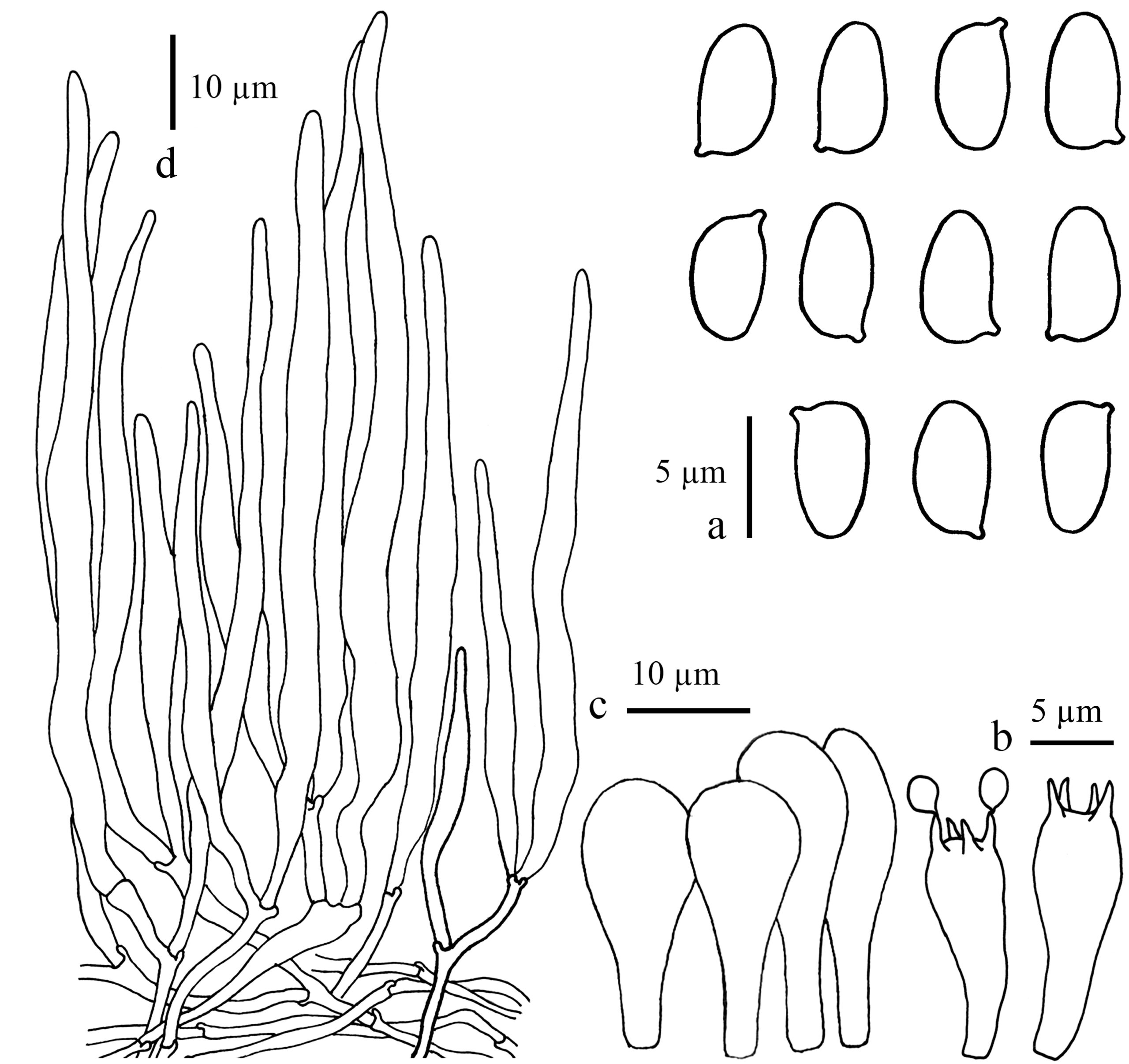
Fig. 2 – Microcharaters of Lepiota venenata (HNL503248). a Basidiospores. b Cheilocystidia. c Elements structure on pileus covering.
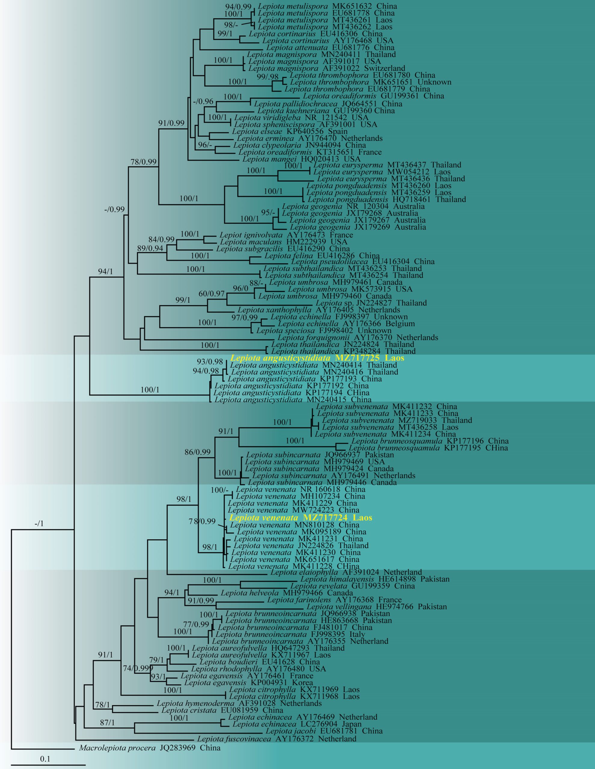
Fig. 3 – Maximum likelihood phylogenetic tree (ML) of Lepiota based on nrITS sequences. One hundred and eight strains consisting of 683 characters after alignment (including gaps). The tree topology derived from the Bayesian analysis was similar to that derived from the ML analysis. The best RaxML tree with a final likelihood value of -11952.807294 is presented. The matrix had 474 distinct alignment patterns, with 7.95% undetermined characters or gaps. Evolutionary model applied is GTRGAMMA. Bootstrap values of ML equal to greater than 70% and Baysian posterior probabilities equal to greater than 0.94 are given above branches. The tree is rooted with Macrolepiota procera (JZB2115036). The newly generated sequence is indicated in yellow.