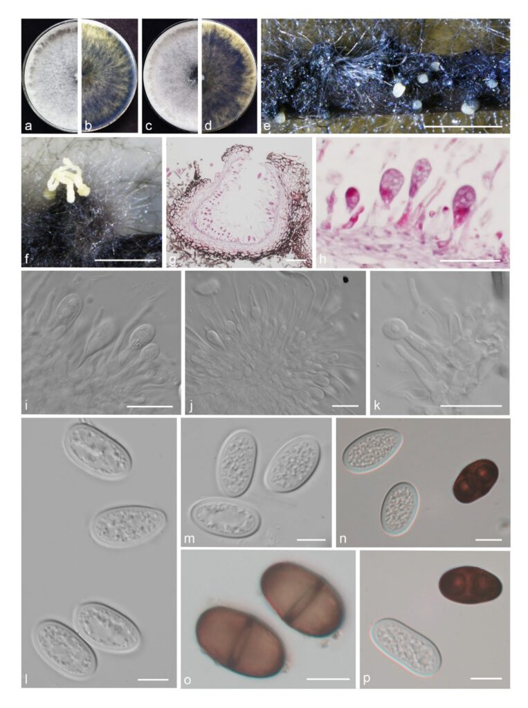
Lasiodiplodia xinyangensis Yanfen Wang & M. Zhang, sp. nov. Fig. 21
MycoBank number: MB 843698; Index Fungorum number: IF 843698; Facesoffungi number: FoF 12935;
Etymology: Referring to the city, Xinyang, where it was collected.
Holotype: HMAS 351941
Description
The sexual stage was not observed. Conidiomata pycnidial, produced on pine needles on WA with 2–3 wk, solitary or aggregated, globose to subglobose, immersed or semiimmersed in the substrate, black, up to 240 μm high, up to 420 μm wide, releasing in saffron yellow to pale yellow conidial tendrils or mass. Paraphyses filiform, hyaline, tip rounded, aseptate, not branched, up to 66 μm in length, 2–3 μm wide. Conidiophores reduced to conidiogenous cells. Conidiogenous cells hyaline, cylindrical, terminal, straight, (8.5–)9–12.5(–14) × (3–)3.5–4(–4.5) μm. Conidia (22–)23–25(–26) × (12–)12.5–13.5(–14.5) μm (av. = 24 × 13 μm, n = 50; L/W = 1.8), initially hyaline and aseptate, ellipsoid to ovoid with rounded or slightly tapered apex, thickwalled (1–2 μm), with granular content, becoming brown and 1-septate, with longitudinal striations.
Material examined: CHINA, Henan Province, Xinyang City, from branch of grapevine, fruiting structures induced on needles of Pinus sp. on water agar, 10 Dec. 2019, Y.F. Wang & M. Zhang (holotype HMAS 351941, culture ex-type CDZM 026 = CGMCC 3.20839); Zhengzhou City, from branch of grapevine, 12 Jun. 2021, Y.F. Wang & M. Zhang (culture CDZM 578).
Sequence data: ITS: OL863156 (ITS1/ITS4); EF1a: OM243825 (728F/986R); TUB2: OM228638 (Bt2A/Bt2B); RPB2: OM243833 (RPB2-LasF/ RPB 2-LasR)
Notes: Lasiodiplodia xinyangensis is introduced based on the multi-locus phylogenetic analysis, with two isolates clustering separately in a well-supported clade (BI/ML = 1/100). L. xinyangensis is most closely related to L. chiangraiensis and L. eriobotryae, but differentiated from them on condia. Conidia of L. xinyangensis (av. = 24 × 13 μm) are smaller than those of L. chiangraiensis (av. = 25 × 14 μm) (Wu et al. 2021) and L. eriobotryae (av. = 26.3 × 14.9 μm) (this study). In addition, conidia of L. xinyangensis quickly become dark brown and longitudinal striations, while the conidia of L. chiangraiensis are hyaline without longitudinal striations. In terms of the nucleotide comparison, L. xinyangensis differed from these species by nucleotide differences in tef1 (L. chiangraiensis: 7 bp, L. eriobotryae: 7 bp), tub2 (L. chiangraiensis: 2 bp) and rpb2 loci (rpb2 is not available to L. chiangraiensis, L. eriobotryae: 5 bp).

Fig. 21 Lasiodiplodia xinyangensis. a–d. colony growing on front and back of PDA (a, b) and MEA (c, d); e–f. conidiomata on PNA; g–h. section view of conidiomata; i–k. conidiogenous cells and developing conidia between paraphyses; l–m. hyaline conidia; n, p. hyaline, aseptate and brown, mature conidia; o. brown, 1-septate mature conidia. — Scale bars: e–f = 500 μm; g = 50 μm; h, j = 20 μm; i, k–p = 10 μm.
