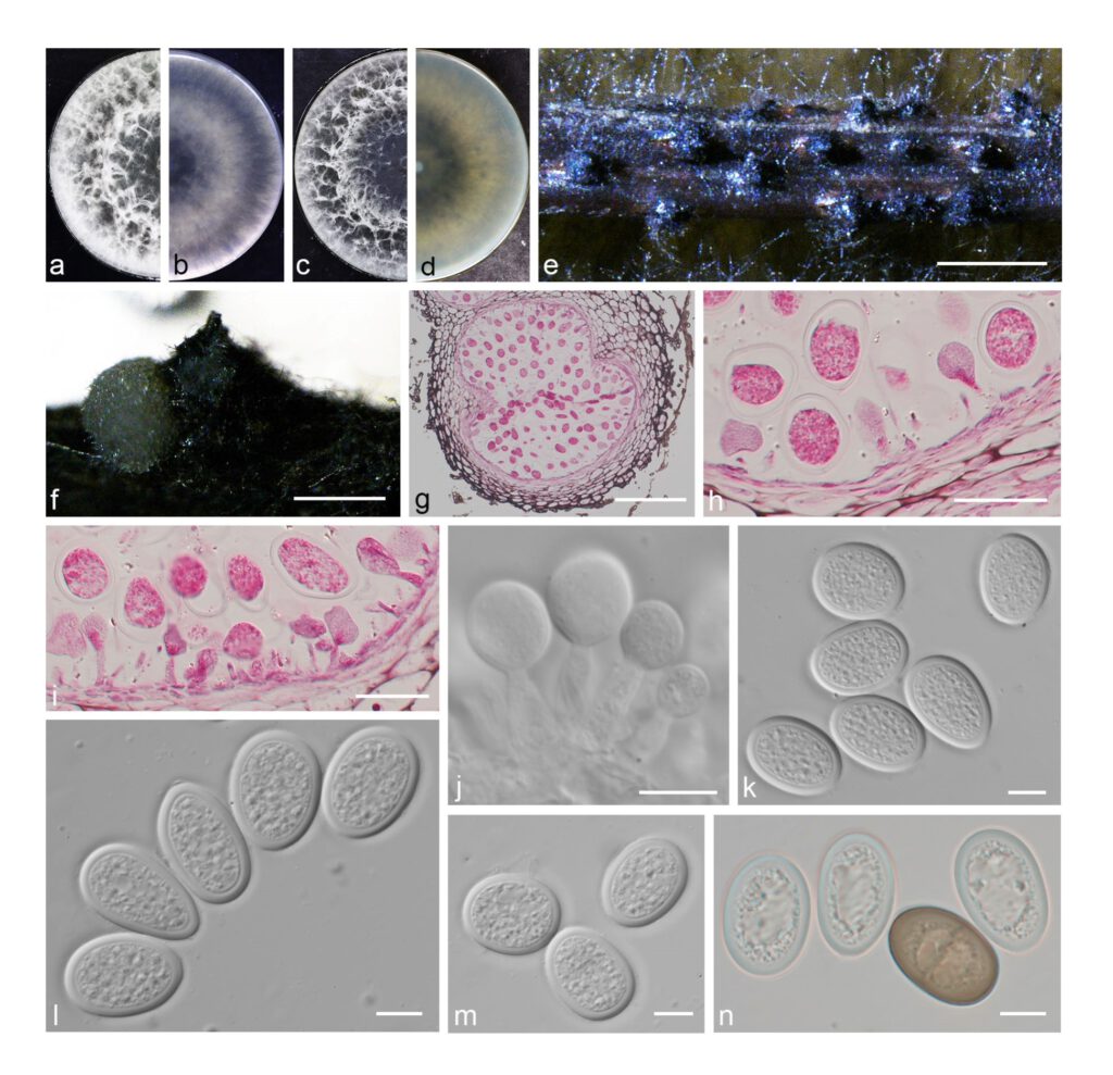
Lasiodiplodia ziziphi Yanfen Wang & M. Zhang, sp. nov. Fig. 22
MycoBank number: MB 843626; Index Fungorum number: IF 843626; Facesoffungi number: FoF 12936;
Etymology: Name refers to zizyphus jujuba, the host from which this fungus was collected.
Holotype: HMAS 351911
Description
The sexual stage was not observed. Conidiomata pycnidial, produced on pine needles on WA with 2–3 wk, mostly uniloculate, black, solitary, globose with a central ostiole, superficial or rarely semiimmersed, up to 270 μm high, up to 610 μm wide, releasing in pale yellow conidial mass. Conidiophores reduced to conidiogenous cells. Conidiogenous cells hyaline, smooth, holoblastic, proliferating percurrently, cylindrical, sometimes slightly swollen at the base, (8–)9.5–12 × (3–)3.5–4 μm. Conidia initially hyaline, subglobose to oval, apex rounded, widest in middle to upper third, with granular content, wall < 2 μm, remaining so for a long time, turning brown with a median septum and vertical striations when mature, (22–)24–27.5(–29) × (15.5–)16.5–19.5(–20) μm (av. = 25.7 × 18.1 μm, n = 50; L/W = 1.4).
Material examined: CHINA, Henan Province, Zhengzhou City, from branches of Jujube, fruiting structures induced on needles of Pinus sp. on water agar, 10 May 2019, Y.F. Wang & M. Zhang (holotype HMAS 351911, culture ex-type CDZM 004 = CGMCC 3.20838); ibid., culture CDZM 005.
Sequence data: ITS: OL863173 (ITS1/ITS4); EF1a: OM243826 (728F/986R); TUB2: OM228639 (Bt2A/Bt2B); RPB2: OM243828 (RPB2-LasF/ RPB 2-LasR)
Notes: Lasiodiplodia ziziphi forms a well-supported, independent clade in Lasiodiplodia. L. ziziphi is closely related to L. crassispora. But it can be distinguished morphologically based on the conidial dimensions. Conidia of L. ziziphi (av. = 25.7 × 18.1 μm) are shorter and wider than those of L. crassispora (av. = 28.8 × 16.0 μm) (Treena et al. 2006). Furthermore, in terms of nucleotides, on ITS it differs two nucleotides, five in the tef1, one in the tub2 and five in the rpb2 from L. crassispora.

Fig. 22 Lasiodiplodia ziziphi. a–d. colony growing on front and back of PDA (a, b) and MEA (c, d); e–f. conidiomata on PNA; g–i. section view of conidiomata; j. conidiogenous cells and developing conidia; k–m. hyaline conidia; n. hyaline, aseptate and brown, mature conidia. — Scale bars: e = 500 μm; f = 200 μm; g = 100 μm; h–i = 20 μm; j–n = 10 μm.
