Kirschsteiniothelia acutisporum S. Wang, Q. Zhao & K.D. Hyde sp. nov.
MycoBank number: MB; Index Fungorum number: IF; Facesoffungi number: FoF 11799; Fig. ***
Etymology:
Holotype: X
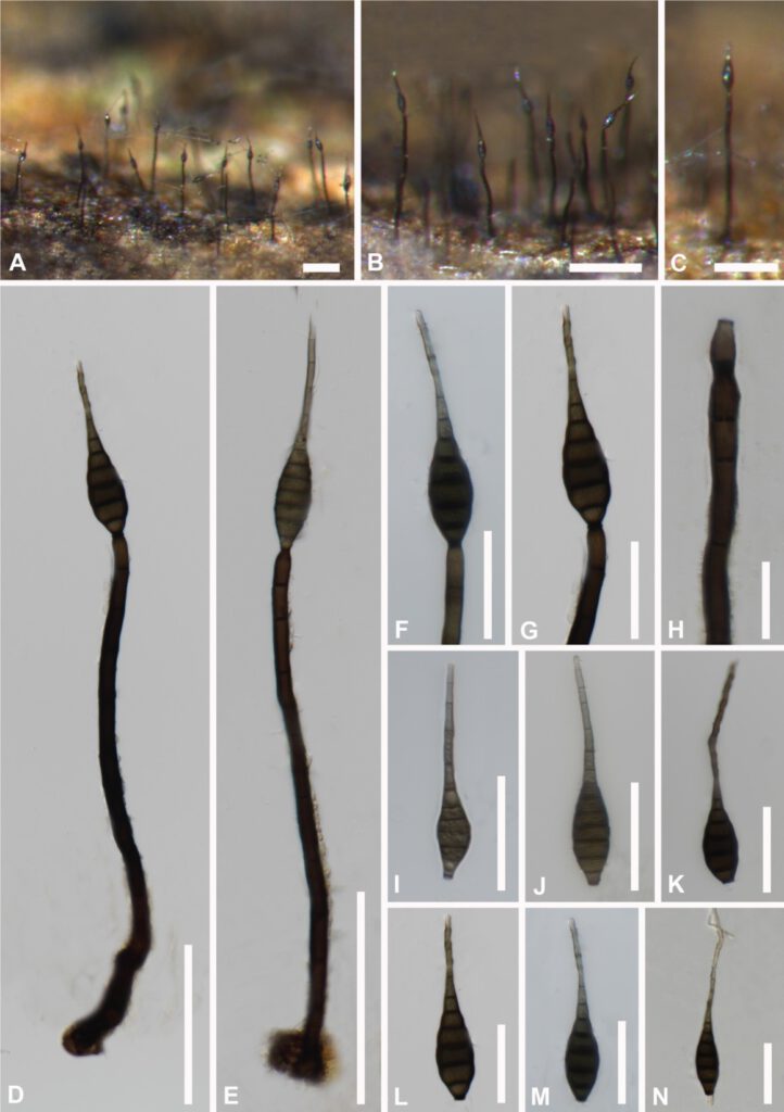
Saprobicon decaying plant substrates. Sexual morph: Not observed. Asexual morph: Colonies effuse, scattered, dark brown to black, hairy, glistening. Mycelium partly superficial, partly immersed in the substratum, composed of brown, septate, branched hyphae. Conidiophores macronematous, mononematous, solitary, cylindrical, straight or slightly flexuous, dark brown, slightly tapering towards the apex, 8–12 septate, truncate at the apex, 180–260 µm (x̄ = 230 µm, n = 10) long, 7–12.5 μm (x̄ =9 µm, n = 10) wide. Conidiogenous cells integrated, terminal, monoblastic, cylindrical and\, brown, calyciform. Conidia acrogenous, solitary, obclavate to obspathulate, tapering to the apex, rostrate, 7–12-euseptate, mid to dark brown, becoming pale brown to pale towards the apex, truncate at the base, 75–120 µm (x̄ = 92 µm, n = 15) long, 10.5–19.5 μm (x̄ = 15 µm, n = 15) wide.
Culture characteristics: Conidia germinating on PDA within 24h. at room temperature. Colonies on PDA, regular round shape, becoming filamentous in the culture, mycelium slightly raised, flattened, filiform, gray aerial hyphae, spreading from the center, becoming olivaceous-brown at the surface and olivaceous-brown to black in reverse, PDA change to light gray to olivaceous-brown (Fig. * ).
Material examined: Thailand, Chiangmai Province, saprobic on decaying wood in MRC, August 2020, Song Wang, SW231, (MFLU 21-0127, holotype), extype culture, *****.
Notes: Kirschsteiniothelia acutisporum shares similar characters with K. fluminicola in having macronematous, unbranched, cylindrical, septate, conidiophores and solitary, obclavate, septate, conidia. However, Kirschsteiniothelia acutisporum differs from K. fluminicola in having a gelatinous rounded sheath at the apex of shorter and thinner conidia (33–43 × 7.5–8.5 μm vs 47.5–86.5 × 8–10μm).

Fig ** Kirschsteiniothelia acutisporum (MFLU 21-0127, holotype) A–C Colonies on dead wood. D,E Conidiophore with conidia. F,G Conidiogenous cells and conidia. H Conidiogenous cell. I–M Conidia. N Germinating conidium. Scale bars: A = 100 μm, B = 200 μm, C–E = 100 μm, F,G= 50 μm, H = 20 μm, I–N = 50 μm.
