Kirschsteiniothelia septemseptatm S. Wang, Q. Zhao & K.D. Hyde sp. nov.
MycoBank number: MB; Index Fungorum number: IF; Facesoffungi number: FoF 11800; Fig. 2
Etymology: Referring to the collection site from Chiang Rai Province in Thailand.
Holotype: MFLU 21-0126
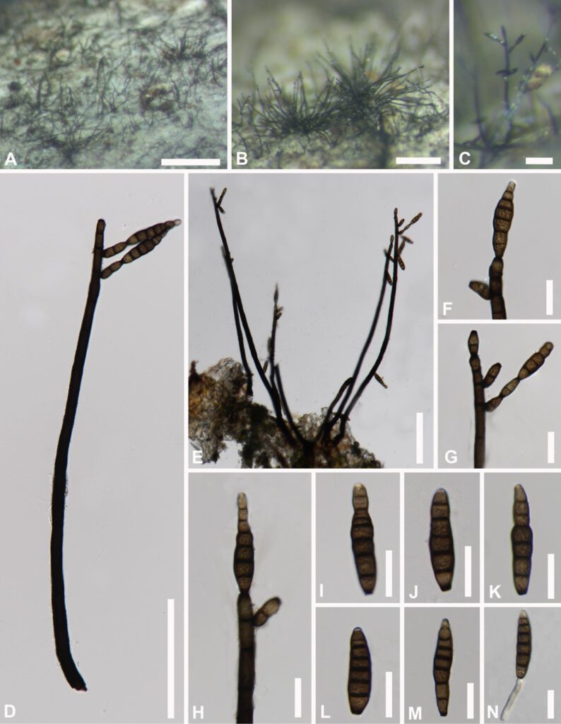
Saprobic on decaying wood. Sexual morph: Not oberved. Asexual morph: Colonies on natural substrate, scattered or fascicular, effuse, hairy, dark brown to black, glistening. Mycelium partly superficial, partly immersed in the host tissue, composed of smooth, light brown, branched, septate. Conidiophores macronematous, mononematous, single to loosely fasciculate, erect, straight to slightly flexuous, branched at the apex, dark brown, multiseptate, 9–16 septate, 250–580 µm (x̄ = 415 µm, n = 20) long, 6.5–14.5 μm (x̄ = 10 µm, n = 20) wide. Conidiogenous cells mostly polytretic, sometimes monotretic, integrated, discrete, terminal and lateral, calyciform, 2 septate, 9.5–21 µm (x̄ = 16 µm, n = 20) long, 4–8 μm (x̄ = 6 µm, n = 20) wide. Conidia acrogenous, solitary, dry, olivaceous brown to brown, pale at apex, obclavate, rostrate, smooth, straight or curved, truncate at base, 5–8– euseptate, 25–55 μm (x̄ = 41 µm, n = 20) long, 6.5–12.5 µm (x̄ = 10.5 μm , n = 20) wide.
Culture characteristics: Conidia germinating on PDA within 24hrs at room temperature. Colonies on PDA irregular, mycelium slightly raised, moderately fluffy, filiform, olivaceous-brown aerial hyphae at the surface, spreading from the center and dark brown to olivaceous-brown in reverse from the center with light brown at rim. (Fig. *).
Material examined: Thailand, Chiangmai Province, saprobic on decaying wood in MRC, July 2020, Song Wang, SW212, (MFLU 21-0126, holotype), ex-type culture, MFLUCC ****.
Genbank numbers:
Notes: Kirschsteiniothelia septemseptatm shares similar characters with K. fluminicola in having macronematous, unbranched, cylindrical, septate, conidiophores and solitary, obclavate, septate, conidia. However, K. cangshanensis differs from K. fluminicola in having a gelatinous rounded sheath at the apex of shorter and thinner conidia (33–43 × 7.5–8.5 μm vs 47.5–86.5 × 8–10μm).

Fig * Kirschsteiniothelia septemseptatm (MFLU ****, holotype) A–C Colonies on dead wood. D, E Conidiophore with conidia. F–H Conidiogenous cells and conidia. I–M Conidia. N Germinating conidium. Scale bars: A = 500 μm, B = 200 μm, C = 50 μm, D,E= 100 μm, F–N= 20 μm.
