Isthmomyces Z. F. Yu, M. Qiao & R. F. Castañeda, gen. nov.
MycoBank number: MB 556126; Index Fungorum number: IF 556126; Facesoffungi number: FoF 05740;
Etymology: Latin, isthmus, Greek (isthmós, “neck”) meaning a narrow cellular structure that connectingtwo larger bodies or cells, +Greek, myces referring to fungus.
Sexual morph: undetermined.
Asexual morph: hyphomycetous.
Colonies effuse, pale mouse grey to dark mouse grey. Mycelium superficial and immersed. Conidiophores macronematous, mononematous, erect, unbranched, smooth, pale brown or hyaline, septate, sometimes reduced to conidiogenous cells. Conidiogenous cells poly-blastic, denticulate, integrated, terminal, sympodial extended. Conidial secession schizolytic. Conidia acroge- nous, isthmospore, composed two cellular isthmic-segment obclavate, clavate, pyriform, obpyriform, lageniform, subulate fusiform to navicular to lanceolate, unicellular or septate, smooth, hyaline, connected by a very narrow, distinct or inconspicuous isthmus.
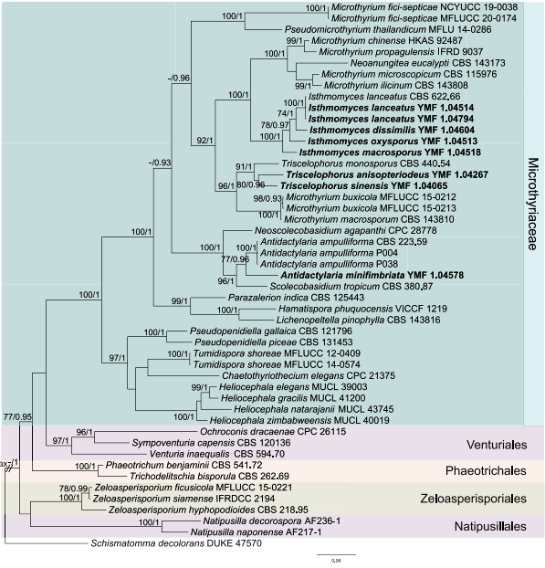
Notes: – Isthmolongispora Matsush. was established with I. intermedia Matsush. as type species [63]. The genus is characterized denticulate, sympodially extending conidiogenous cells and conidia composed by two or several cellular structures, which are connected by very narrow isthmuses. In present study, isthmospore with two and more cellular isthmic-segment specimens were collected respectively. Phylogenetic analysis inferred from four loci showed that the species with isthmospore composed by two cellular isthmic-segment (hemi-isthmospore) belong to Microthyriaceae (Figure 1), while species composed by more than two cellular isthmic- segments belong to Leotiomycetes based on phylogenetic analysis inferred from LSU (Figure 2). We retained species with 3(–4) cellular isthmic-segments which were similar to type species Isthmolongispora intermedia in Isthmolongispora, and established Isthmomyces to comprise two cellular isthmic-segments (hemi-isthmospore).
Type species: Isthmomyces oxysporus Z.F. Yu, M. Qiao & R.F. Castañeda.

Figure 1. Phylogenetic tree generated by the Maximum Likelihood (ML) analysis using combined se- quences of the nuclear large subunit (LSU) and the internal transcribed spacers (ITS) gene. Bootstrap support values for ML over 70% and Bayesian posterior probabilities greater than 0.9 are indicated above or below the nodes as MLBP/BIPP. Schismatomma decolorans strain DUKE 47570 is used as the outgroup. Novel species are indicated in bold.

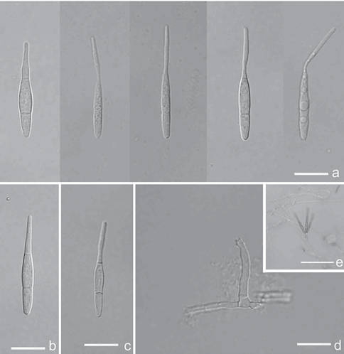
Figure 2. Antidactylaria minifimbriata (Holotype YMF 1.04578) a–c conidia d conidiophore and co- nidiogenous cell e conidia on conidiophore under low objective. Scale bars: 10 μm (a–d); 50 μm (e).
Species
