Hohenbuehelia tristis G. Stev., Kew Bull. 19(1): 26 (1964)
MycoBank number: MB 332010; Index Fungorum number: IF 332010; Facesoffungi number: FoF 10710; Figure 6–7
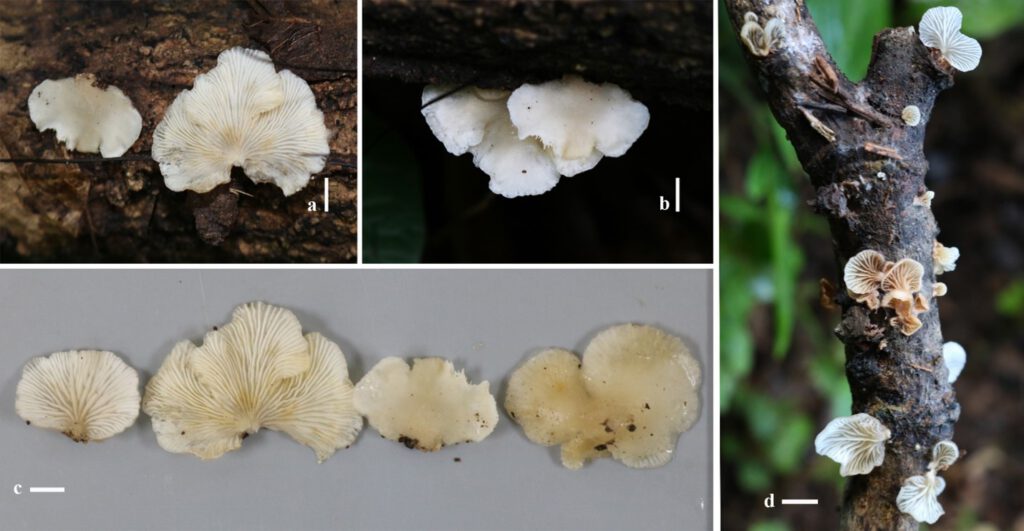
Pileus 20–30 × 15–20 mm, spathuliform to rounded flabelliform, dimidiate to orbicular, laterally or dorsally attached without stipe sometimes present pseudostipe when young then disappeared when old, greyish-white, creamy white (1A1–1B1), plane, upper surface when young appearing tiny fibrillose grayish hair (5A1–5B1) dense at the base and thinly toward margin observed in the lens becoming whitish with age, finally disappeared when a mature stage, glutinous when wet, very faintly translucent, shiny, white (1A1) finely grayish towards base, striate margins. Lamellae radiating from point of attachment, 1 mm thick, very crowded, white to dirty white (1A1). Order and Test not observed. Spore print white.
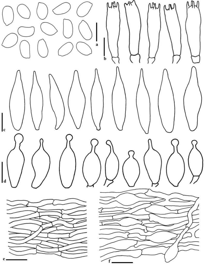
Basidiospores (4.6–)5.4–6.6–7.7(–8.1) × (3–)3.1–3.9–4.8(–5.1), Q = (1.21–)1.34–1.37–2.12(–2.14), ellipsoid to sub ellipsoid, smooth, inamyloid, thin-walled. Basidia (13–)13.9–21.2–23.9(–24.2) × (3.5–)5.5–6.6(–7) µm, clavate to subcylindraceous, with 4 sterigmata, 2–4 µm long, 4-spored or sometimes 2-spored, hyaline, smooth, thin-walled. Hymenophoral trama irregular, made up of gelatinized hyphae up to 9.5 µm wide, hyaline. Cheilocystidia (10.9–)10.9–13.1–18.7(–19.8) × (4.2–)4.4–5.5–6.5(–6.5) µm, lecythiform to sub lageniform with absent or one swelling protruding from the apical portion, with a neck and capitulum. Pleurometuloids present in both side of lamellar (37.9–)38.3–61.5–82(–82.3) × (10.7–) 10.9–15–18.5(–18.6) µm, narrowly fusiform, fusiform to subfusiform with a narrow base, mostly with a lanceolate apical part and partly, brownish, thick welled covered with encrusted with crystals observed in water, not present in KOH 5 %. Context consisting of gelatinized zone with compound hyphae 1.40–2.55 µm wide, thick as revived in 5 % KOH. Hymenophoral trama subregular. Pileipellis observed on young stage present tufts of a trichoderm and mature stage present an ixocutis of variously intertwined, slightly gelatinized, filamentous, smooth hyphae, 3–9.5 µm wide, with a light brown intracellular pigment, with undifferentiated terminal elements. Pileus trama 50–450 µm deep, made up of hyphae up to 9 µm wide and with walls up to 1 µm thick. Clamp connections are present ubiquitously.
Habitat: Solitary, gregarious to imbricate, growth on dead small branches.
Specimens examined: Nang Lae Nai village, Muaeng district, Chiang Rai on 31st July 2019, Monthien Phonemany (MT85 and MT86).
Notes: Hohenbuehelia tristis from Thailand has characterized by small creamy white basidiomata covered with tiny finely fibrillose hair then disappeared with mature stage and later glutinous when wet, very faintly translucent, shiny. Hohenbuehelia tristis is characterized by small creamy white basidiomata, glutinous when wet, covered with tiny finely fibrillose hair, very faintly translucent and shiny, disappearing when matured. The morphological description of our specimens fits well with the description of the holotype of H. tristis, originally described from New Zealand. But H. tristis has larger metuloids 80–90 × 15–20 µm, pseudo-amyloid, pileipellis covered by tufts of parallel thin-walled (Stevenson 1964). Our study was observed both young, mature and old basidiomies, where for example pileipellis were observed in the different stages. In our specimens’ collections the fibrillose hair on the pileipellis when young disappeared as the basidiomes matured, therefore the pileipellis is present as both a trichoderm and an ixocutis.
According to phylogenetic analysis in Fig. 1, H. grisea from Thailand (as culture MFLUCC 12-0451) and H. grisea (HFJAU0029) from China (Unpublished) clustered together with our collection of H. tristis with high bootstrap supported (BS). Both Thai and Chinese sequence of H. grisea from Genbank have no morphological descriptions as evidence. Therefore, these might be wrongly identified as they are 100% similar to our collection of H. tristis. Additional confirmations was made by comparing the morphology of our specimens with H.grisea which was originally described as Pleurotus atrocoeruleus var. griseus Peck from New York. It is distinguished by a greyish-black, sparsely tomentose pileus, the lamellae becoming cream-colored when aged, partially gelatinous (New York State Museum 1890). H. tristis (RV95/214 DUKE) by Moncalvo et al. (2000) was identified base on LSU sequence only which 0.99% identities from our specimen and the phylogeny tree H. trisis from Australia in Table 1, is closed related with our specimen with high BS.

Fig. 6: Basidiomata of Hohenbuehelia tristis in the field (a,b,c=MT85 (MFLUXXX); d=MT86 (MFLUXXX); scale bar 1 cm).

Fig. 7: Micro-morphological of Hohenbuehelia tristis. a = Basidiospores, b = Basidia, c = Pleurometuloids, d = Cheilocystidia, e =Context hyphae, f = Pilieipellis (scale bar a–b = 10 µm, c = 20 µm, d–f = 10 µm).
