Halojulella avicenniae (Borse) Suetrong, K.D. Hyde & E.B.G. Jones, Phytotaxa 130(1): 19 (2013). Fig. 83
MycoBank number: MB 803343; Index Fungorum number: IF 803343; Facesoffungi number: FoF 06533.
Saprobic on decaying wood of Avicennia marina, frequently young twigs. Sexual morph: Ascomata 200–400 high × 235–360 µm diam. (x̅ = 305 × 285 µm, n = 10), globose or subglobose, immersed beneath a clypeus, membranous, with ostiolate. Ostiole 100–150 long × 80–100 µm diam. (x̅ = 118 × 91 µm, n = 5), periphysate. Peridium 2-layered, 20–40 µm (x̅ = 28 µm, n = 5), thickened above with clypeal tissue, outer layer of small pseudoparenchymatous cells, brown, inner layer of hyaline cells. Hamathecium comprising cellular, hypha-like, septate pseudoparaphyses. Asci 125–195 × 175–30 µm, (x̅ = 195 × 20 µm, n = 15), 8-spored, bitunicate, fissitunicate, thick- walled, clavate, moderately long pedicel with club-like base, apically rounded, with an ocular chamber surrounded by distinct apical apparatus, not bluing in IKI (I-), developing from the base of the ascoma. Ascospores 27–40 × 12.5–15 µm, (x̅ = 35 × 13 µm, n = 25), overlapping 1–2-seriate, ellipsoidal, hyaline, with a central septum when young, becoming yellow to pale brown, or golden brown, with 6–7 transsepta when mature, constricted particularly at the central septum with up to 2–3 longisepta, and surrounded by a large spreading sheath. Asexual morph: Coelomycetous, phoma-like. Conidiophores 10–25 × 2–3 µm, (x̅ = 17.5 × 2.7 µm, n = 10), filiform, septate, branched; Conidia 1.5–5 × 2–4 µm, (x̅ = 3.5 × 2.8 µm, n = 15), globose to ellipsoidal, hyaline, aseptate, thin-walled.
Culture characteristics – Ascospores germinating on 2 % sea water agar within 24 h with germ tubes produced from both ends. Colonies on malt extract sea water agar initially yellow when mature turns into brown, reverse brown, reaching 15 to 30mm in diameter in 25 days at room temperature. Mycelium hyaline to brown, producing yellow brown pigments, velvety.
Material examined – India, Tamil Nadu, Pondicherry, Veerampattinam mangroves, (11.59°N 79.5°E), on decaying wood of Avicennia marina (Acanthaceae), 28 November 2015, B. Devadatha, AMH-9996, living culture, NFCCI-4424.
GenBank numbers – ITS: MK028713, LSU: MK026757, rpb-2: MN532682, SSU: MK026754, tef1: MN532686.
Notes – Our specimen is identical to the type species Halojulella avicenniae (Fig. 83). The asexual morph of our species was found on media and it produced conidiophore and conidia from culture, while fruiting body is not observed (Fig. 84). Phylogenetic analyses (Fig. 42) support our species as H. avicenniae by grouping with other strains of H. avicenniae with high support (100 % MLBS, 100 PP).
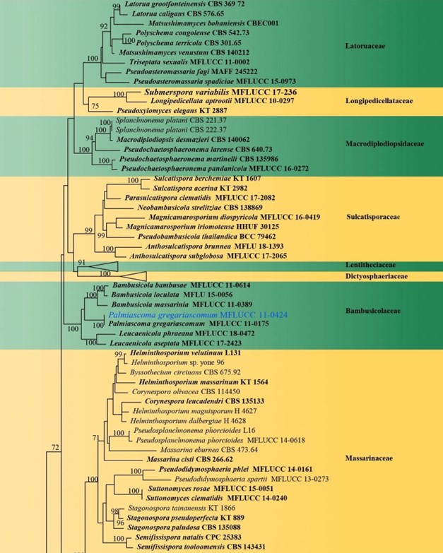
Figure 42 – Phylogram generated from maximum likelihood analysis (RAxML) of Pleosporales based on ITS, LSU, rpb-2, SSU and tef1 sequence data. Maximum likelihood bootstrap values equal or above 70 % are given at the nodes. An original isolate number is noted after the species name. The tree is rooted to Capnodium coffeae (CBS 147.52). The ex-type strains are indicated in bold.

Figure 42 – Continued.
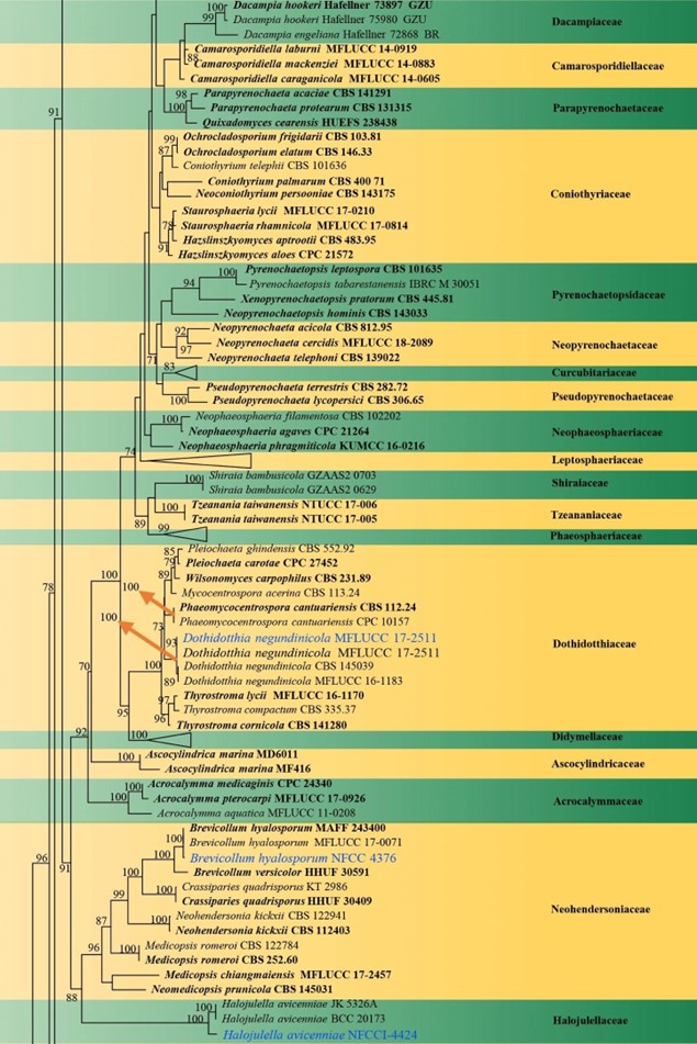
Figure 42 – Continued.
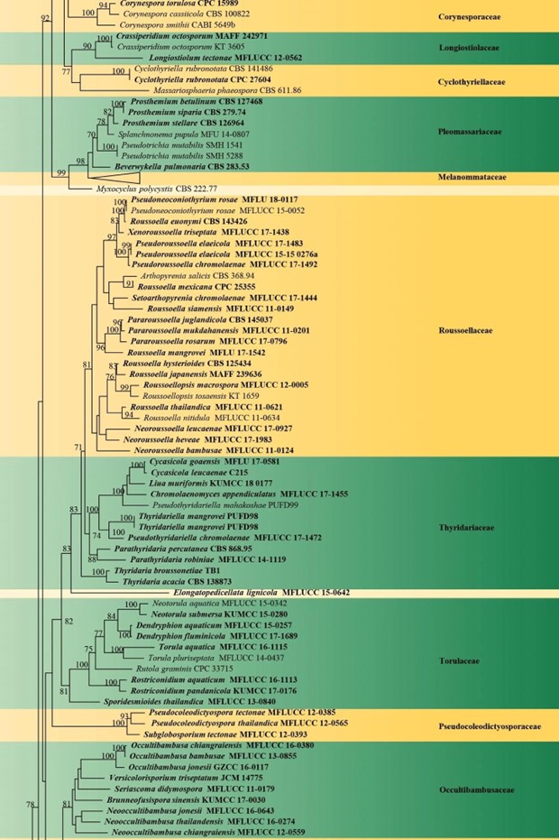
Figure 42 – Continued.
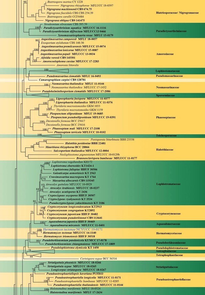
Figure 42 – Continued.
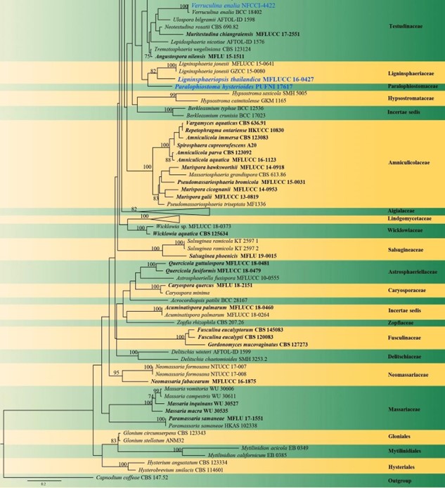
Figure 42 – Continued.
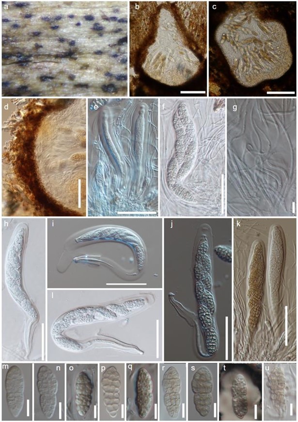
Figure 83 – Halojulella avicenniae (AMH-9996). a Ascomata erumpent on decaying wood. b, c Longitudinal sections of ascomata. d Section of peridium. e, f, h Immature asci. g filamentous pseudoparaphyses. i, l Asci showing fissitunicate dehiscence. j, k Mature asci. m–s Hyaline to brown ascospores. t Wide gelatinous sheath in India ink. u Germ tubes developed from terminal ends of ascospore. Scale bars: b, c = 100 μm, d–f, h–k, = 50 μm g, m–u = 10 μm.
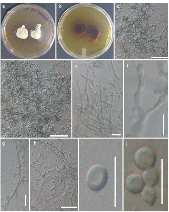
Figure 84 – Halojulella avicenniae (AMH-9996). a–b Culture. c–h Stages of conidiophore bearing conidia from culture. i–j Conidia. Scale bars: c, d, h, = 50 μm e, f, i–j = 10 μm.
