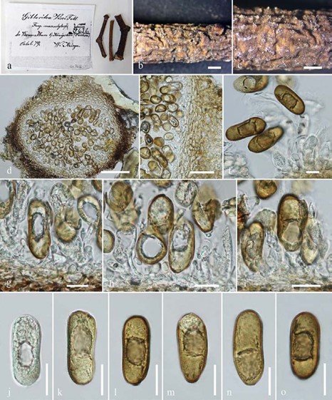
Gibberidea visci Fuckel, Jb. nassau. Ver. Naturk. 23-24: 168 (1870) [1869-70] Fig. 3
Current name: Phaeobotryosphaeria visci (Kalchbr.) A.J.L. Phillips & Crous, in Phillips, Alves, Pennycook, Johnston, Ramaley, Akulov & Crous, Persoonia 21: 47 (2008)
MycoBank number: MB 227688; Index Fungorum number: IF 227688; Facesoffungi number: FoF 06243;
Saprobic or pathogenic on undetermined plant stem. Sexual morph: See Phillips et al. (2013). Asexual morph: Conidiomata 290–397 μm high × 422−454 µm diam. ( x = 327.3 × 444.8 µm, n = 10), immersed to erumpent, unilocular, subglobose. Peridium 33−55 µm composed of dark brown cells of textura angularis. Conidiogenous cells 4–13 µm × 7−14 µm ( x = 8.5 × 10.8 µm, n = 10), hyaline, enteroblastic, discrete, proliferating internally to form periclinal thickenings. Conidia 34– 43 μm × 14−17 µm ( x = 40.1 × 16.7 µm, n = 10), oval, apex obtuse to rounded, base obtuse or truncate, moderately thick-walled, initially hyaline, becoming brown, externally smooth-walled, internally finely verruculose.
Notes – Phillips et al. (2008) recorded the size of conidiomata of Sphaeropsis visci as ‘up to 300 µm’, conidiogenous cells as ‘(4–) 8.5–11× 4–6.5 µm’ and conidia as ‘(27–29–33(–50) × (14.5–) 15.5–19(–22) µm’ which is similar to our examined specimen.
Material examined – GERMANY, Saxony, on submerged decaying leaves, 7 November 1965, J.L. Crane (ILL00088985).
Economic significance – Sphaeropsis visci is a pathogen of Viscum album (mistletoe) (Varga et al. 2012).

Figure 3 – Gibberidea visci (ILL 00088985) a, b Herbarium material. c Appearance of conidiomata on host surface. d Section of conidioma. e Peridium. f–i Conidiogenesis. j–o Conidia. Scale bars: b, c = 1 mm, d = 100 μm, e–o = 20 μm.
