Gamsomyces Hern.-Restr. & R´eblov´a, gen. nov.
Index Fungorum number: MB 834446; Facesoffungi number: FoF 13280
Etymology: This genus is named in honour of the late Walter Gams, our colleague and friend, for his contribution to mycology.
Type species: Gamsomyces longisporus (M.B. Ellis) Hern.-Restr. & R´eblov´a
Description: Asexual morph: Colonies with sporodochial or synnematous conidiomata. Conidiophores semi-macronematous or macronematous, fasciculate, simple or penicillately branched, subhyaline to brown. Conidiogenous cells monoblastic, inte- grated, terminal, elongating percurrently. Conidia dry, solitary, curved, fusiform, pigmented, transversely euseptate, with a mucilaginous cap at the apex. Conidia secede schizolytically.
Sexual morph: unknown.
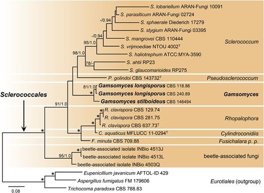
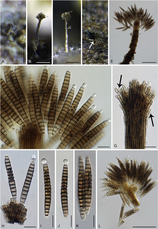
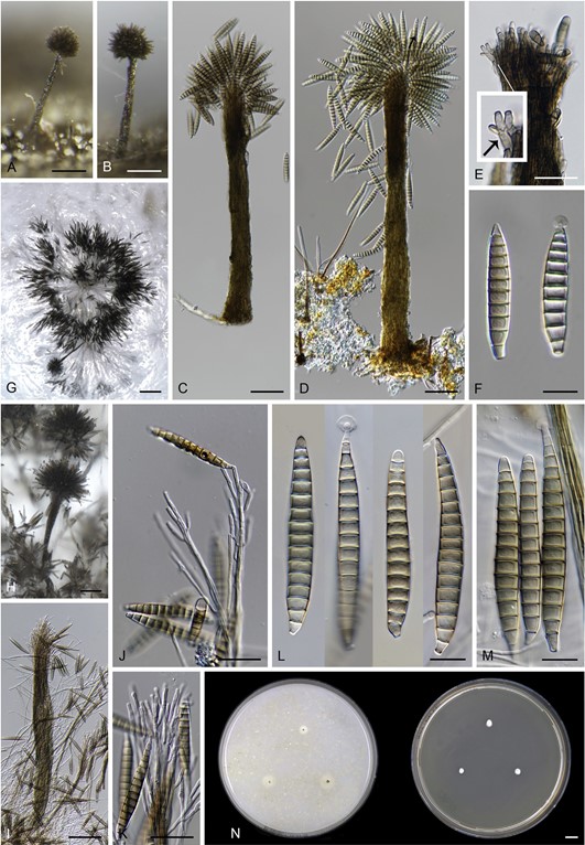
Notes: – Multigene phylogenetic analysis of three strains of Bac- trodesmium longisporum and B. stilboideum revealed they were unrelated to Bactrodesmium; they formed a strongly supported lineage in the Sclerococcales (Eurotiomycetes), which is intro- duced as the new genus Gamsomyces (Fig. 4). Two species are accepted in the genus, and new combinations are proposed. Gamsomyces differs from Bactrodesmium in having inconspic- uous, brown to olivaceous brown conidiomata, both sporodochia and synnemata, presence of a mucilaginous cap at the apex of conidia, absence of dark bands over the transverse septa and morphology of the conidiogenous cells. The percurrently elon- gating conidiogenous cells on the natural substrate, first mentioned by Hughes (1978) in G. longisporus, are in agreement with our observations (Fig. 15G). The same mode of the con- idiogenous cell elongation is also present in G. stilboideus (Fig. 17E).
Key to species of Gamsomyces
1a. Sporodochia and synnemata on the natural substrate, synnemata 74–305 μm long, conidia 11–16-septate,
48.5–74 × 6– 8 μm, or longer up to 80 μm fide (Ellis 1976) and 95 μm fide (Hughes 1978) with up to 21 septa; in vitro only sporodochia formed, conidia 41– 163.5 × 5.5–8 μm, (6–)14–25-septate…………………………………………… G. longisporus
1b. Only synnemata 380 – 455 μm long on the natural sub- strate, conidia 10– 13-septate, 46– 69 × 7– 9 μm, or shorter 30 – 55 × 7– 8 μm (fide Casta~neda-Ruiz & Arnold
1985); in vitro synnemata 325 – 633 μm long, conidia 42– 90 × 6.5 – 10 μm, (5–)14 – 16-septate…G. stilboideus

Fig. 4. Combined phylogeny using ITS, LSU, SSU and rpb2 of representatives of the Sclerococcales. Species names given in bold are taxonomic novelties, T indicates ex-type strains. An asterisk (*) indicates branches with ML BS = 100 %, PP values = 1.0. Branch support of nodes ≥70 % ML BS and ≥0.90 PP is indicated above or below branches.

Fig. 15. Gamsomyces longisporus. A–C. Synnemata (A, viewed from the top). D. Sporodochium-like conidioma (indicated by arrow). E, L. Upper part of the synnema with conidiogenous cells and conidia. F, H. Conidia and conidiogenous cells. G. Conidiogenous cells (arrows indicate percurrently elongating conidiogenous cells. I–K. Conidia. A– L. On natural substrate. Images: A, K, L CBS H-3931, B– J CBS H-3972. Bars: A– D = 100 μm, E, L = 50 μm, F– K = 10 μm.

Fig. 17. Gamsomyces stilboideus (CBS 146494). A– D. Synnemata. E. Upper part of the synnema with conidiogenous cells (arrow indicates percurrently elongating con- idiogenous cells). F, L, M. Conidia. G, H. Synnemata and densely branched conidiophores with conidia. J, K. Conidiophores with conidia. A–F. On natural substrate. G– M. On CBSOA. N. Colonies on CBSOA and MEA after 2 wk. Bars: A, B, H = 100 μm, C, D, I = 50 μm, E, J, K = 25 μm, F, L, M = 10 μm, G = 200 μm, N = 1 cm.
Species
