Fusiformispora clematidis Phukhams., M.V. de Bult & K.D. Hyde, in Phukhamsakda et al., Fungal Diversity: 10.1007/s13225-020-00448-4, [12] (2020)
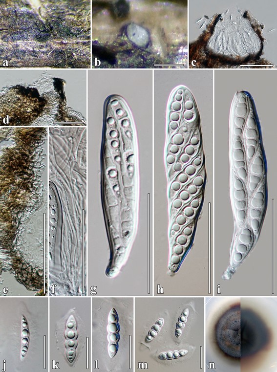
MycoBank number: MB 557107; Index Fungorum number: IF 557107; Facesoffungi number: FoF 07243, Fig. 3.
Etymology: Epithet reflects the host Clematis.
Holotype: MFLU 17–1485.
Saprobic on dead stems of Clematis fulvicoma. Sexual morph: Ascomata 165–190 × 200–275 μm (x̄= 175 × 225 μm, n = 5), on the surface of the host, covered by a pseudoclypeus, visible as black spots, immersed to superficial, solitary, scattered, uniloculate, obpyriform to compressed globose, base flattened, brown to dark brown, partially carbonaceous, rough- walled, with apical ostioles. Ostioles central, 55 × 35 μm, brown to dark brown, papillate, with easy opening by a pore, filled with periphyses. Peridium 10–18 μm wide, multi-layered, comprising 4–5 layers of brown to dark brown cells of textura angularis, inner layers comprising thin, hyaline cells. Hamathecium composed of dense, 0.5–1.5 μm wide (x̄= 1.3 μm, n = 50), filiform, branched, trabeculate pseudoparaphyses, anastomosing above the asci, reaching the ostiole, transversely septate. Asci 86–127 × 18–24 μm (x̄ = 100 × 25 μm, n = 40), 8-spored, bitunicate, fissitunicate, thick-walled, cylindric-clavate, apically rounded, short, with furcate pedicel, ocular chamber clearly visible when immature. Ascospores 24–36 × 5–10 μm (x̄ = 30 × 8 μm, n = 50), biseriate, partially overlapping, broadly fusiform, tapering towards the ends, acute at both ends, hyaline, with (1–)3–4 transverse septa, with large guttules in each cell, constricted at the septa, deeply constricted at the median septum, cell above median septum slightly wider than below, smooth- walled, with 4–12 μm wide mucilaginous sheath. Asexual morph: Undetermined.
Culture characters: Colonies on MEA reaching 50 mm diam. after 4 weeks at 25 °C. Cultures from above, grey- brown, with reddish brown mixed in the mycelium, dense, colonies circular, flat, umbonate, raised from the agar in the centre, dull, covered with aerial mycelium, white mycelium at the edge; reverse dark brown, dense, circular, with irregular, fimbriate margin, pinkish mycelium radiating outwardly. Material examined: Thailand, Chiang Rai Province, on dead stems of Clematis fulvicoma Rehder & E.H. Wilson, 20 March 2017, C. Phukhamsakda, CMTH22 (MFLU 17–1485,
holotype); ex-type living culture, MFLUCC 17–2077.
Host: Clematis fulvicoma—(This study).
Distribution: Thailand—(This study).
GenBank accession numbers: LSU: MT214542; SSU: MT226661; ITS: MT310589; tef1: MT394725; rpb2: MT394677.
Notes: In a BLASTn search of GenBank, the LSU sequence of Fusiformispora clematidis MFLUCC 17–2077 showed 96% similarity to Lindgomyces pseudomadisonensis KT 2742 (LC149916), while the ITS sequence had 91% similarity to Vargamyces aquaticus CBS 636.91 (NR_154471). Fusiformispora clematidis is phylogenetically distinct, therefore, we introduce the collection as a new species.

Fig. 3 Fusiformispora clematidis (MFLU 17–1485, holotype). a Appearance of ascoma on host surface. b Close up of ascoma on host substrate. c Vertical section of ascoma. d Ostiolar canal. e Section of peridium. f Pseudoparaphyses. g–i Asci. j–m Ascospores. n Culture characteristics on MEA. Scale bars: b = 200 µm, c = 100 µm, d, j–m = 20 µm, e–i = 50 µm
