Sclerenchymomyces clematidis Phukhams. & K.D. Hyde, sp. nov.
MycoBank number: MB 557111; Index Fungorum number: IF 557111; Facesoffungi number: FoF 07288, Fig. 24.
Etymology: Name refers to the host plant, Clematis.
Holotype: MFLU 16–2492.
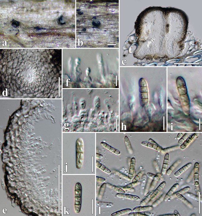
Saprobic on dead stems of Clematis vitalba. Sexual morph: Undetermined. Asexual morph: Conidiomata 187–225 × 85–217 μm ( x̄= 210 × 130 μm, n = 10), pyc-nidial, solitary, sometimes aggregated, uniloculate or multiloculate, erumpent or superficial on host substrate, with black shiny ostioles visible, globose to subglobose,
coriaceous, dark brown to brown, ostiolate. Ostioles central, papillate, oblong. Conidiomatal wall 14–30(–35) μm wide, multilayered, scleroplectenchymatous cells, flat at base, outer layer composed of 5–9 layers of light brown to brown cells of textura angularis, lined with a thick hyaline layer bearing conidiogenous cells. Conidiophores reduced to conidiogenous cells. Conidiogenous cells 3–8 × 1.5–4 μm ( x̄ = 5 × 3 μm, n = 30), enteroblastic, phialidic, determinate, discrete, sub-cylindrical to truncate, smooth-walled, hyaline, arising from the inner layers of conidiomata. Conidia 11–18 × 2.5–5 μm (x̄ = 14 × 4 μm, n = 50), broad cylindrical to oblong, rounded at both ends, hyaline when immature, yellowish at maturity, slightly curved, 3-septate, with 1(–2) guttules in each cell, smooth-walled.
Culture characters: Colonies on MEA reaching 40 mm diam. after 4 weeks at 25 °C. Cultures from above, cream- brown, spare mycelia, circular, umbonate, papillate with fluffy, covered with white aerial mycelium; reverse dark brown at the centre, cream radiating outwardly.
Material examined: Italy, Forlì-Cesena Province, near Meldola, on dead aerial branch of Clematis vitalba, 15 November 2013, E. Camporesi, 1518C (MFLU 16–2492, holotype); ex-type living culture, MFLUCC 17–2180.
Host: Clematis vitalba—(This study).
Distribution: Italy—(This study).
GenBank accession numbers: LSU: MT214558; SSU: MT226675; ITS: MT310605; tef1: MT394737; rpb2: MT394686.
Notes: Sclerenchymomyces clematidis is distinct from S. jonesii in conidial characters (Fig. 24). Sclerenchymomyces clematidis has broad cylindrical to oblong, yellowish conidia with 3 septa and 1(–2) guttules, while S. jonesii has hyaline, aseptate conidia (Wanasinghe et al. 2016a). In a BLASTn search of GenBank, the LSU sequence of S. clematidis (strain MFLUCC 17–2180) was 98.7% similar to S. jonesii (≡ Neoleptosphaeria jonesii), while the ITS sequence showed 97.28% similarity to NR_152375. Pairwise compari- son of the ITS sequence reveals nine bases pair differences (1.59%) between S. clematidis and S. jonesii (MFLUCC 16–1442). The tef1 region shows seven bases pair difference between S. clematidis and S. jonesii.

Fig. 24 Sclerenchymomyces clematidis (MFLU 16–2492, holotype). a Appearance of conidiomata on Clematis vitalba. b Close up of conidioma on host substrate. c Vertical section through conidiomata. D Ostiole from above. e Section of conidioma wall. f–i Conidiogenous cells and conidia. j–l Conidia. Scale bars: b = 200 µm, c = 100 µm, d = 20 µm, e = 50 µm, f–l = 5 µm
