Eremomyces bilateralis Malloch & Cain, Can. J. Bot. 49(6): 849 (1971).
MycoBank number: MB 313945; Index Fungorum number: IF 313945; Facesoffungi number: FoF 05361; Fig. 34
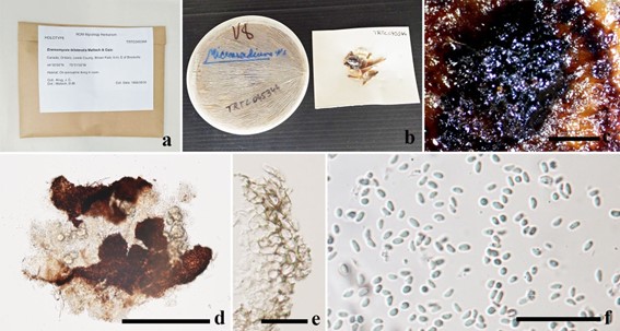
Saprobic on dung of the North American porcupine. Colonies flocculent, drift white to dark brown, superficial, compact, and growing slowly on agar media. Hyphae septate, branched, hyaline to brown. Sexual morph: Ascomata solitary, scattered, superficial on hyphae, globose to ellipsoid, dark brown to black, lacking ostioles. Peridium thin, composed of large brown cells of textura angularis. Hamathecium lacking pseudoparaphyses. Asci 8-spored, bitunicate, obovoid, thin-walled, pedicellate, evanescent. Ascospores multi-seriate, fabiform, hyaline, aseptate, smooth-walled (adapted from Hyde et al. 2013). Asexual morph: Coelomycetous. Pycnidia in culture brown to dark-brown, globose to subglobose, thin-walled, papillate. Conidiophores filiform, septate, hyaline to brown. Conidiogenous cells formed inside the swollen part. Conidia 3.2–4.8 × 1.7–2.8 μm (x̄ = 3.85 × 2.3 μm, n = 20), ellipsoidal, hyaline, aseptate, thin-walled, guttulate.
Material examined: Canada, Ontario, Leeds Co., East of Brockville, on porcupine dung in cave, 5 September 1966,J.C. Krug (TRTC 45344, holotype).
Notes: Eremomyces bilateralis is found on dung of usually sedentary animals, particularly rodents in Canada (Ontario), Kenya, Tanzania and the USA (California). Colonies are relatively slow growing on most media. Ascomata tend to be fragmented when immature and lack characteristic cephalothecoid peridium when mature. On the natural substrate the ascomata are equally conspicuous and produce short setae compared to long hairs seen in culture.

Fig. 34 Eremomyces bilateralis (TRTC 045344, holotype). a Herbarium packet and label. b Hebarium culture c Pycnidium on the upper surface of agar media. d Squash mount of pycnidium. e Conidia. Scale bars: c = 2000 µm, d = 300 µm, e = 25 µm, f = 50 µm
Species
