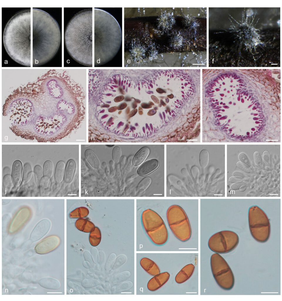
Dothiorella chaenomelina Yanfen Wang & M. Zhang, sp. nov. Fig. 13
MycoBank number: MB 843701; Index Fungorum number: IF 843701; Facesoffungi number: FoF 12932;
Etymology: Name refers to Chaenomeles sinensis, the host from which this fungus was collected.
Holotype: HMAS351944
Description
The sexual stage was not observed. Conidiomata pycnidial, produced on pine needles on WA within 2–3 wk, solitary or aggregated, superficial or semi-immersed, globose to subglobose, covered with hyphal hairs, dark brown to black, thick-walled, uni- to multi-loculate, papillate with a central ostiole, up to 372 μm high, up to 321 μm wide. Conidiophores absent. Conidiogenous cells (7.5–)8–10(–11)×2.5–3.5(–4.5) μm, cylindrical to lageniform, discrete or integrated, unbranched, holoblastic, proliferating at the same level giving rise to periclinal thickenings, aseptate, hyaline, smooth. Conidia oblong to ellipsoidal, initially hyaline, thin-walled, aseptate, becoming brown, 1-septate, occasionally slightly constricted at septum, moderately thick-walled, smooth, ends rounded, often with a truncate base, (16–)16.5–19(–20.5) × (7–)8.5–10.5(–12) μm (av. = 17.8 × 9.8 μm, n = 50; L/W =1.8).
Material examined: CHINA, Henan Province, Zhengzhou City, from branch of pawpaw, fruiting structures induced on needles of Pinus sp. on water agar, 12 Jul. 2021, Y.F. Wang & M. Zhang (holotype HMAS 351944, culture ex-type CDZM 398 = CGMCC 3.20836); Zhengzhou City, from branch of jujube, 12 Jul. 2021, Y.F. Wang & M. Zhang (culture CDZM 241); Beijing City, from branch of plum, 7 Oct. 2020, Y.F. Wang & M. Zhang (culture CDZM 460).
Sequence data: ITS: OL863147 (ITS1/ITS4); EF1a: OM228652 (728F/986R); TUB2: OM228621 (Bt2A/Bt2B)
Notes: The three isolates studied form a well-supported independent clade distinct from known Dothiorella species. Dothiorella chaenomelina is phylogenetically (Fig. 13) closely related to Do. plurivora and Do. acericola, but can be distinguished from them by the size of conidia. Do. chaenomelina have (av. =17.8 × 9.8 μm) smaller conidia than Do. plurivora (17.8 × 9.8 vs 22.5 × 11 μm) (Abdollahzadeh et al. 2014) and Do. acericola (17.8 × 9.8 vs 20.8 × 9.2 μm) (Phookamsak et al. 2019). In addition, Do. chaenomelina colonies cover the 90 mm plates after 3 d, while Do. acericola reaches 70–73 mm after 1 week (Phookamsak et al. 2019), Do. plurivora reaches 62–84 mm after 4 d (Abdollahzadeh et al. 2014). This species differed by nucleotide sequences in ITS (Do. plurivora: 3 indel, Do. acericola: 1 indel), tef1 (Do. plurivora: 1 bp, Do. acericola: 5 bp), and tub2 (Do. plurivora: 2 bp and 2 indel, Do. acericola: 2 bp and 2 indel).

Fig. 13 Dothiorella chaenomelina. a–d. colony growing on front and back of PDA (a, b) and MEA (c, d); e–f. conidiomata on PNA; g–i. section view of conidiomata; j–m. conidiogenous cells and developing conidia; n–o. conidiogenous cells and brown, mature conidia; p–r. brown, 1-septate mature conidia. — Scale bars: e = 500 μm; f = 100 μm; g = 50 μm; h–i = 20 μm; j–m = 10 μm.
