Didymocyrtidium Vain., Acta Soc. Fauna Flora fenn. 49(no. 2): 228 (1921)
MycoBank number: MB 1553; Index Fungorum number: IF 1553; Facesoffungi number: FoF 09047;
Saprobic on growing stem of unidentified trees. Sexual morph: Ascomata semi immersed to superficial on host surface, simple, visible as dark brown to black circle on host surface. Peridium thick-walled, composed of several layers of dark-brown cells of textura angularis. Hamathecium present, with cellular, branching, pseudoparaphyses, anastomosing, branching, embedded in mucilaginous matrix, sometimes aparaphysate, covering asci by gelatinous matrix. Asci bitunicate, fissitunicate, 8-spored, ovoid to saccate, sessile with indistinct ocular chamber, thick-walled. Ascospores crowded, or irregularly arranged in the ascus, oblong or oval to ellipsoid, hyaline to subhyaline 1-septate, very slightly constricted at the septum, smooth or rough-walled. Asexual morph: Undetermined.
Type species – Didymocyrtidium nudum Vain.
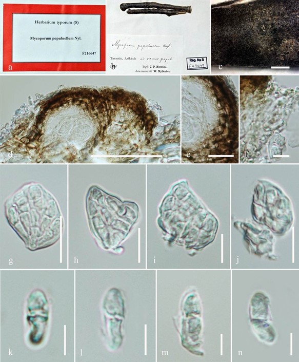
Notes – The genus Didymocyrtidium was introduced by Vainio (1921) to accommodate three species namely Didymocyrtidium mozambicum Vain, Didymocyrtidium nudum and Didymocyrtidium populnellum. The major morphological characters of the genus are semi- immersed to superficial ascomata and unique subhyaline or pale greenish, 1-septate ascospores. The asexual morph is unknown. Cultures and sequences are unavailable. No type species was assigned to this genus. In the original description, the authors described Didymocyrtidium populnellum as a synonym of Mycoporum populnellum Nyl. However, according to index fungorum, Mycoporum populnellum is accommodated in its own family Mycoporaceae. We re- examined the holotype specimen of Didymocyrtidium populnellum (= Mycoporum populnellum) under the code F216647 from S herbarium. Upon examination of the specimen, we observed two fungi. One of them (Fig. 47) is characterised by semi-immersed to superficial simple, black ascomata on host surface with ovoid to saccate asci and hyaline to subhyaline 1-septate ascospores
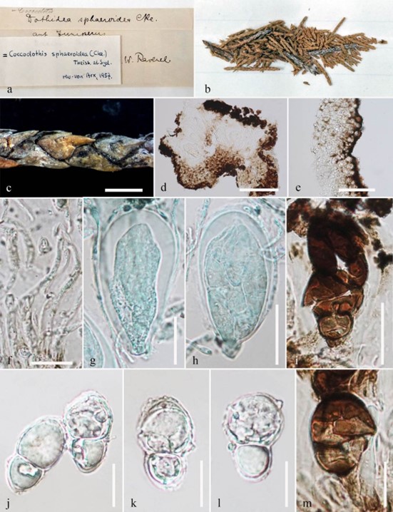
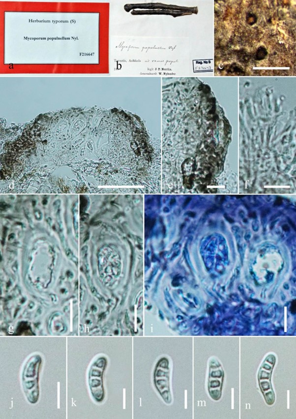
which match the description of Didymocyrtidium populnellum as given by Vainio (1921), while the other fungus (Fig. 48) is characterised by semi-immersed to superficial apothecia on its host surface, uni-loculate, forming pseudostroma or clypeus, 4-spored bitunicate asci with subhyaline or pale greenish, 3–4-septate ascospores which seems to be Mycoporum populnellum. The ascopores of Mycoporum populnellum were illustrated as 3–4-septate in a work by Millot (1899), while Nylander (1873) described the ascospores of M. populnellum as 1-septate. Hence, we cannot typify the genus Didymocyrtidium with the above specimen. We studied the syntype specimen of Didymocyrtidium nudum Vain. from H herbarium under the code H6022676 and illustrate the characters (Fig 42). Hence, in this study we typify the genus Didymocyrtidium with the type specimen Didymocyrtidium nudum. We also report that Didymocyrtidium populnellum and Mycoporum populnellum are two different taxa. We provide an updated illustration for Didymocyrtidium populnellum, Mycoporum populnellum and Didymocyrtidium nudum. We retain the genus Didymocyrtidium in Dothideomycetes, genera incertae sedis until sequence data becomes available.

Figure 42 – Coccodothis sphaeroidea (S-F51270, isotype). a, b Details of herbarium material. c Appearance of ascomata on host surface. d Section of ascoma e Peridium. f Hamathecium. g–i Asci. j–m Ascospores. Scale bars: c = 500 µm, d = 100 µm, e, g–h = 20 µm, f, j–m = 10 µm.

Figure 47 – Didymocyrtidium populnellum (S-F216647, holotype). a Details of herbarium material. b, c Habit and appearance of ascomata on host surface. d Section of ascoma. e Peridium. f Hamathecium. g–j Asci. k–n Ascospores. Scale bars: c = 2 mm, d = 50 µm, e–j = 10 µm, k–n = 5 µm.

Figure 48 – Mycoporum populnellum (S-F216647, holotype). a Details of herbarium material. b, c Habit and appearance of ascomata on host surface. d Section of ascoma. e Peridium. f Hamathecium. g–i Asci. j–n Ascospores. Scale bars: b = 2 mm, c = 1 mm, d = 50 µm, e–i = 10 µm, f, j–n = 5 µm.
Species
