Diaporthe beilharziae R.G. Shivas, Jacq. Edwards & Y.P. Tan, in Tan, Edwards, Grice & Shivas, Fungal Diversity 61: 254 (2013)
Index Fungorum number: IF 802383, Facesoffungi number: FoF 00013
Etymology – In recognition of Dr Vyrna Beilharz, a highly respected Australian mycologist who first collected and isolated this fungus.
Conidiomata pycnidial, solitary, scattered or aggregated in small groups, abundant on PDA, OMA, and wheat straw pieces on WA after 4 weeks, solitary and immersed in WA after 4 weeks, subglobose, up to 250 μm diam, ostiolate, beaks absent or less than 300 μm, abundant pale yellow to salmon conidial droplets exuded from ostioles; walls thin, composed of an inner layer of yellowish-brown textura angularis and an outer layer of darker yellowish brown textura epidermoidea. Conidiophores formed from the inner layer of the locular wall, reduced to conidiogenous cells or 1-septate, hyaline to pale yellowish brown, ampulliform to cylindrical, 5–15 × 1.5–3.5 μm, Conidiogenous cells cylindrical to flexuous, tapered towards the apex, hyaline, 5–20 × 1.5–3.0 μm. Alpha conidia abundant, oval to cylindrical, rounded at the apex, obconically truncate at base, mostly biguttulate, hyaline, (5.5–) 6.5–9 (−10) × 2–2.5 (−3) μm. Beta conidia scarce amongst the alpha conidia, flexuous, hyaline, 15–25 × 1.0–1.5 μm. Perithecia not seen.
Cultural characteristics — Colonies on PDA covering the entire plate after 10 days, adpressed to slightly ropey with pycnidia visible as hundreds of small black dots, transparent becoming pale greyish sepia towards the centre; reverse similar to the surface. On OMA covering the entire plate after 10 days, adpressed, transparent to pale mouse grey with pycnidia apparent as small black dots or irregular patches less than 200 μm diam.; reverse similar to the surface.
Specimens examined — Australia, New South Wales, Mittagong, on Indigofera australis, 30 April 1991, V.C. Beilharz; holotype VPRI 16602 (includes ex-type culture); isotype BRIP 54792.
Notes — Diaporthe beilharziae was isolated from a leaf spot on Indigofera australis. Two other species, Diaporthe indigoferae on dead branches of I. gerardiana in Pakistan (Müller and Ahmad 1958) and P. indigoferae on stems of I. dosua and I. dalea from Europe (Uecker 1988), have been reported on Indigofera. Conidia were not described for D. indigoferae. Phomopsis indigoferae has larger alpha conidia (8 × 3–4 μm) than D. beilharziae (5.5–10 × 2–3 μm). The role of all of these fungi as pathogens is not known. The phylogenetic inference from the combined sequence data showed D. beilharziae clustered close to D. infecunda (Gomes et al. 2013). In culture, D. beilharzae produced abundant pycnidia on PDA and OMA, compared to D. infecunda, which was sterile.
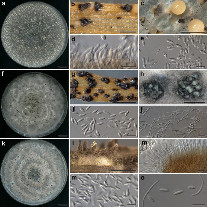
Fig. 1 – Diaporthe beilharziae (ex-type BRIP 54792) after 4 weeks, a culture on PDA, b pycnidia on sterilised wheat straw, c conidial ooze, d conidiophores, e alpha conidia and a solitary beta conidium. Diaporthe fraxini–angustifoliae (ex-type BRIP 54781) after 4 weeks, f culture on PDA, g pycnidia on sterilised wheat straw, h conidial ooze, i alpha conidia, j beta conidia. Diaporthe litchicola (ex-type BRIP 54900) after 4 weeks, k culture on PDA, l pycnidia on sterilised wheat straw, m conidiophores, n alpha conidia, o alpha and beta conidia. Scale bars: a, f, k = 1 cm; b–c, g–h, l = 1 mm; d–e, I–j, m–o = 10 μm
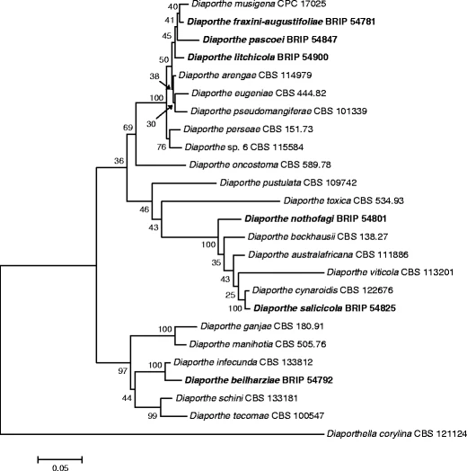
Fig. 2 – Maximum likelihood tree inferred from analysis of three combined three genes (ITS, TEF and BT). The percentages of replicate trees in which the associated taxa clustered together in the bootstrap test (1000 replicates) are shown next to the branches. The tree was rooted to Diaporthella corylina. The alignment and tree are deposited in TreeBASE (S13424). Species described in this study are in bold