Cryptophiale udagawae Piroz. & Ichinoe, in Pirozynski, Can. J. Bot. 46: 1126 (1968)
Index Fungorum Number: IF 329371
Saprobic on decaying wood. Asexual morph: Colonies on plant substrate effuse, hairy, scattered, glistening. Mycelium mostly immersed, and partially superficial. Conidiophores macronematous, mononematous, setiform, scattered, erect, straight or slightly curved, dark brown, smooth, thick-walled, septate below the branches, 150–290 × 10–33 μm, 1–2 times dichotomously branched towards the apex and generally above the fertile region; terminal branches dark brown, 9.5–51.5 μm long, 2.9–6.2 μm wide at the base, acutely pointed above. Fertile region from middle up to the first dichotomy, 36.5–92 × 10–17 μm, consisting of two rows of phialides one on each side of the conidiophore with each cell narrowly ellipsoid, covered by sterile shield cells. Conidiogenous cells not observed. Conidia produced in slime and adhering to the fertile region, hyaline, smooth, clavate, aseptate, basal end rounded, with a short appendage at the base, 19.9–28.1 × 1.6–2.8 μm (av. = 24.9 × 2.05 μm, n = 30). Sexual morph: Undetermined.
Material examined: CHINA, Guizhou Province, Qiannan Buyi Miao Autonomous Prefecture, Dushan County, Guizhou Zilinshan National Forest Park (Shengou District), unnamed road, on decaying wood, 6 July 2018, Chuan-Gen Lin, DS 1-36 (MFLU 19–0209, HKAS 105133), living culture GZCC 18–0047.
Notes: This is the second report of Cryptophiale udagawae from China and we provide description, sequences data and phylogenetic analysis for this species. This collection fits well with the description of Pirozynski (1968) and with the first description of a Chinese isolate by Yang et al. (2018). The phylogenetic result (Fig. 1) showed that our isolate clustered with C. udagawae (MFLUCC 18–0422 and MFLUCC 18–0428) with high support (91% MLBS, 0.95 PP). Thus, we identify our collection as C. udagawae.
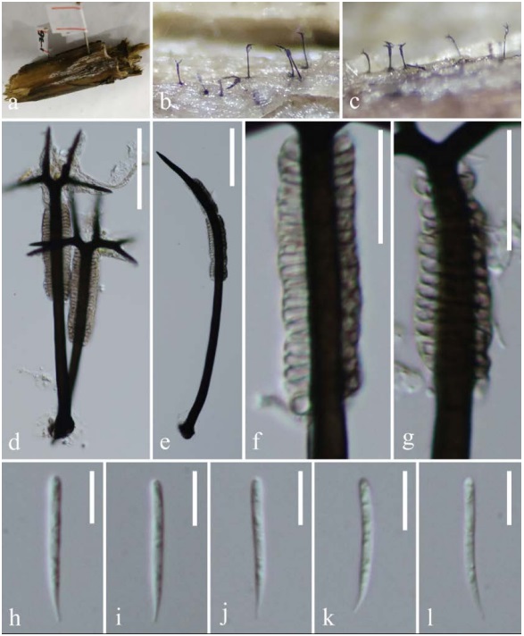
Figure 1 – Cryptophiale udagawae (MFLU 19–0209). a Host material. b, c Conidiophores on the host surface. d, e Conidiophores and conidiogenous cells. f, g Conidiogenous cells. h–l Conidia. Scale bars: d, e = 50 μm, f, g = 20 μm, h–l = 10 μm.