Colletotrichum dioscoricola H. D. Yang & K. D. Hyde, sp. nov.
MycoBank number: MB; Index Fungorum number: IF; Facesoffungi number: FoF 12854;
Description
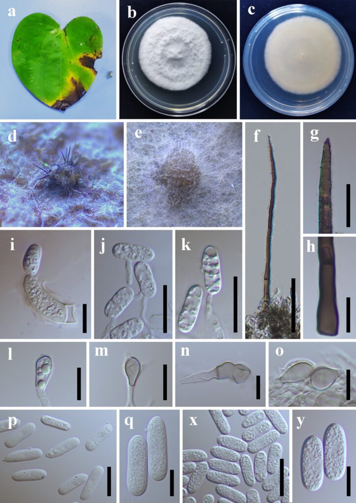
Pathogenic on living leaf of Dioscorea yunnanensis. Sexual morph: undetermined. Conidiomata 332–440 ´ 379–435 μm, acervular, solitary or scattered, semi-immersed, subglobose to globose, or irregularly shape, dark brown, commonly covered by a mass of conidia. Setae 95–160 μm long, 5–7 μm in wide, brown, cylindrical, gradually dwindle toward the upper, 1–8-septate, base smooth-walled, verruculose towards the tip. Conidiophore hyaline, smooth walled. Conidiogenous cells 14–25 × 3.5–6 μm, monophialidic, cylindrical, unbranched, septate. Conidia hyaline, oblong, straight, smooth-walled, aseptate, 20–23 × 6.5–7.7 μm (av. ± SD = 21 ± 1.1 × 6.5 ± 0.3 μm, n = 40), L/W ratio = 3.2.
Colonies on PDA reaching 38–42 mm diam after 10 days, white, flat, cottony, circular, edge entire, reverse white. Conidial masses formed on the surface of medium, abundant, confluent, cream color at beginning, later becoming dark brown when age. Setae not observed. Conidiophores hyaline, smooth-walled, unbranched or branched. Conidiogenous cells hyaline, smooth-walled, periclinal thickening not observed. Conidia hyaline, 0–1-septate, straight, cylindrical, with one obtuse end, 16–21.5 × 6–7.5 (av. ± SD ± 18.3 ± 1.2 × 6.7 ± 0.3, n = 50), L/W ratio = 2.7. Appressoria solitary, light brown to brown, subglobose, clavate, oblong, or irregularly shaped, 5.5–16.5 × 3.8–8.8 μm (av. ± SD = 9.4 ± 2.8 × 6.4 ± 1.4 μm, n = 20). On OA media, reaching 40–46 mm diam after 10 days, white, flat, myceliam dense. edge entire, reverse white with a light brown ring, not pigment, not sporulation. On SNA, vegetative hyphae sparse, hyaline, reaching 30–33 mm diam after 10 days, not sporulation.
Material examined: CHINA, Yunnan, Wenshan, 23°46′39.01″N,104°9′14.98″E, elevation 1536 m, on Dioscorea yunnanensis, October 2021, holotype: HKAS123208, living culture: KUNCC22-10800, KUNCC22-10801.
Distribution: China
Notes: Colletotrichum dioscoricola form a sister species with C. sydowii and C. adenophorae, however, it can be distinguished from C. sydowii in having larger conidia and light brown to brown, subglobose, to oblong appressoria.

Fig1. Colletotrichum dioscoricola. a. Disease symptom on Dioscorea yunnanensis, b. Front and reverse of colony, d. Conidial masses on Dioscorea yunnanensis, e. Conidial masses, f–h. Setae, i. Conidiogenous cells on Dioscorea sp., j–k. Conidiogenous cells, l–o. Appressoria, p–q. Conidia from Dioscorea sp., x–y. Conidia on PDA. Scale bars: i = 10 um, j = 20 um, k = 20 um, f = 20 um, g = 10 um, h = 10 um, l = 10 um, m = 10 um, n = 10 um, p = 20 um, q = 10 um, x = 20 um, y = 10 um.
