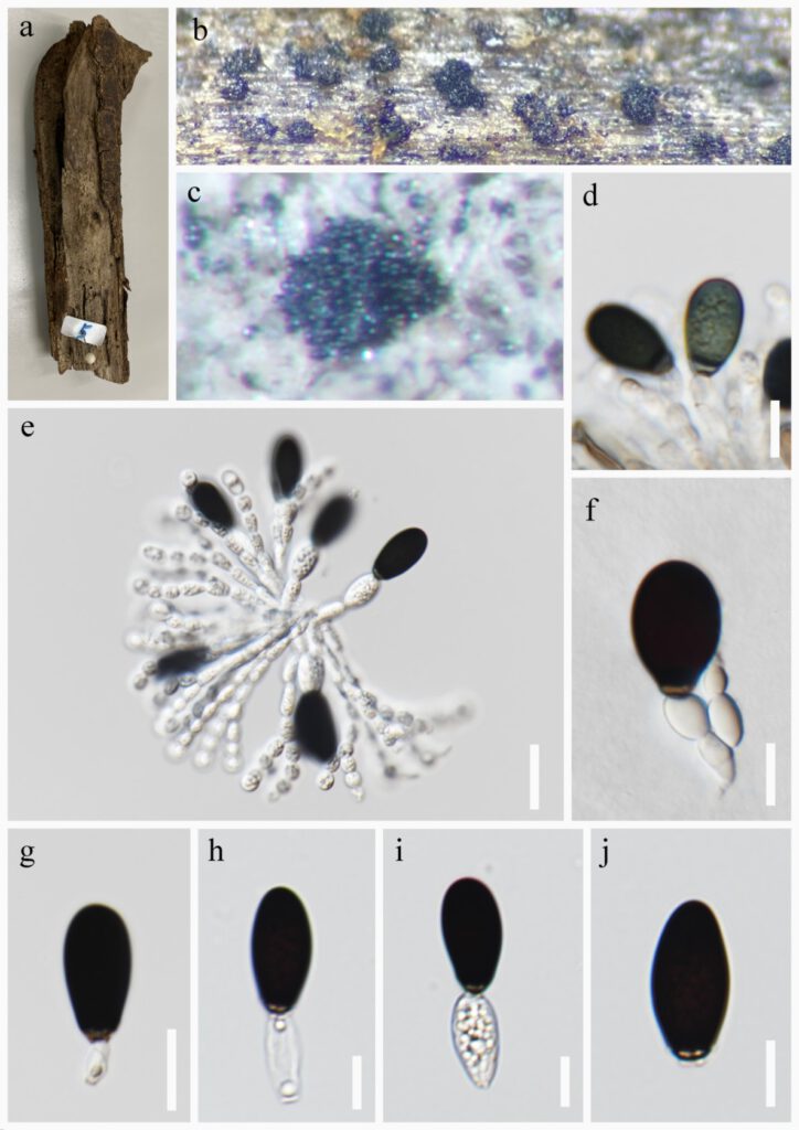
Parafuscosporella lignicola L. Li, R.J. Xu & R. Cheewangkoon sp. nov. FIGURE 3
MycoBank number: MB 559931; Index Fungorum number: IF 559931, Facesoffungi number: FoF 12889
Etymology: Referring to the fungus dwelling on wood.
Holotype:
Saprobic on decaying twigs from freshwater habitats. Asexual morph: Colonies on natural substratum sporodochial, scattered, black without covering. Mycelium mostly superficial, partially immersed, composed of branched, smooth-walled, septate, hyaline to light brown hyphae. Conidiophores semi-macronematous, mononematous, compact, flexuous, simple or branched, smooth-walled, septate, hyaline, with an ellipsoidal cells 12–19 × 6–11 μm (x̅ = 16 × 9 μm, n=15). Conidiogenous cells 11–19 × 7–9 μm (x̅ = 14× 8 μm, n=20) monoblastic, integrate or discrete, terminal, moniliform, ellipsoidal or cylindrical, hyaline, smooth-walled, sometimes with a continuous proliferate. Conidia 15–25 × 8–13 μm (x̅ = 22 × 11 μm, n=40) acrogenous, solitary, subglobose, ellipsoidal or obpyriform, with a septum near the pale brown base, olivaceous when young, dark brown to black when mature, pale brown at basal cell, truncate at base, smooth-walled. Basal cell elliptic, 12–20 × 6–11 μm (x̅ = 16 × 9 μm, n=30). Sexual morph: Undetermined.
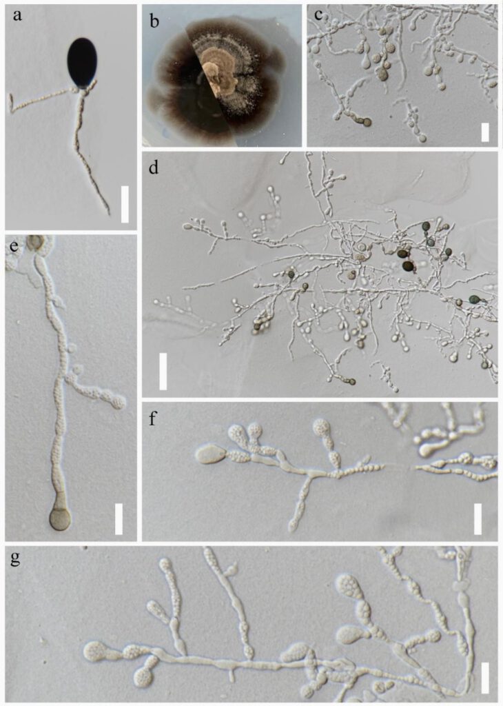
Culture characteristics: Conidia germinating on PDA within 48h. Germ tubes produced from the basal cell. Colony reached 30 mm at room temperature in the dark for 1 month, on PDA media, flat, velutinous, spreading, the colors spread out from the center, the area near the center is light grey, the areas away from the center are brown. With sparse mycelium on the surface, with irregular margin. Conidial formation from single spore germination on PDA. Sporulation appears first in the centre of the colony, later over the whole colony. Conidiophores macronematous, branched, sometimes reduced to a single conidiogenous cell, hyaline to pale brown. Conidiogenous cells monoblastic, holoblastic, integrated, cylindrical, hyaline to pale brown, contracted at the septum. Conidia 10–25 × 8–14 μm (= 17 × 11 μm, n=20), globose to subglobose, olivaceous when young, medium brown to dark brown when mature, aseptate. uniseptate at the base with paler, sometime with a continuous proliferation, cylindrical basal cell 6–15 × 4–7 μm (= 10 × 6 μm, n=20). (FIGURE. 4)
Known distribution: Thailand
Material examined: Nang Lae, Mueang Chiang Rai, Chiang Rai Province, Thailand (99°52′52.93″E, 20°3′2.52″N), Saprobic on decaying wood submerged in freshwater stream, submerged in freshwater stream, 18 July 2020, R.J Xu, MD-5 (MFLU22-0101 holotype), ex-living culture MFLUCC XXXXX. Mushroom Research Center (M.R.C.), Chiang Mai Province, Thailand (98°46′44.28″E, 19°7′7.62N″), unidentified decaying wood in terrestrial habitat, 13 July 2020, R.J Xu, MD-5-3 (MFLU22-0102), living culture MFLUCC XXXXX.
Notes: Morphologically, Parafuscosporella lignicola similar to P. aquatica, P. nilotica, P. moniliformis, P. mucosa, P. obovate and P. pyriformis in having semi-macronematous, mononematous conidiophores; Monoblastic, integrated, globose, subglobose, ellipsoidal conidiogenous cells; Subglobose, ellipsoidal or obpyriform, dark brown to black conidia. However, Parafuscosporella lignicola has moniliform, ellipsoidal conidiophores, ellipsoidal or cylindrical conidiogenous cells and the smallest conidia in the genes (Boonyuen et al. 2016, Yang et al. 2016, 2017, Boonmee et al. 2021).
Phylogenetic analysis also shows that Parafuscosporella lignicola are different other species of Parafuscosporella and form a sister group with P. ellipsoconidiogena (Figure. 2). However, P. ellipsoconidiogena can be distinguished from P. lignicola by having doliiform, fusiform conidiophore, fusiform conidiogenous cell and more big conidia (27.5–33 × 15–20 vs 15–25 × 8–13 μm) (Boonyuen et al. 2021).

FIGURE. 3 Parafuscosporella lignicola (MFLU22-0101 holotype). a Specimen b, c Colonies on substrate. d, e Conidiophores with conidia. f–j Conidiophores, conidiogenous cells and conidia. Scale bars: d = 10, e = 20, f-j = 10 μm.

FIGURE. 4 Sporulation of Parafuscosporella lignicola (XXX ex-type) on PDA. a Germinated conidium on PDA. b Obverse (left) and reverse (right) views of a colony on PDA. c-g Hyphae, Conidiophores, Conidia and conidiogenous cells from culture. Scale bars: a, c = 20 μm, d = 50 μm, e-g =20μm.
