Cladocillium musae Chun-Hao Chen & R. Kirschner, sp. nov. Figs. 2 and 3
MycoBank number: MB 835864; Index Fungorum number: IF 835864; Facesoffungi number: FoF 11139;
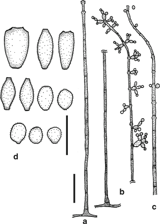
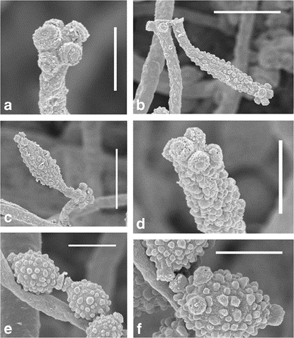
Etymology: Name referring to the host plant genus Musa. Colonies not associated with distinct disease symptoms on the leaf, but with those caused by other fungi, olive-green to brown. Hyphae subhyaline to pale brown, smooth to finely verruculose, 1–2 μm wide. Conidiophores single, erect, with a non-rhizoidal single-celled foot distinguished from vegetative hyphae by deeper pigmentation and broader width, ca. 12–30 μm long and 5–10 μm high, with unbranched stipe (excep- tionally with a single short lateral branch), appearing smooth in light microscopy, occasionally rough in SEM, (43–)87– 222.5(–350) × (2.5–)3–5(–6) μm (n = 30). Distances between septa 14–28 μm, becoming shorter (ca. 10 μm) towards the apex of conidiophore, and cells becoming narrower, 1.5–2 μm, cells of the apical ca. 100–200 μm long part of stipe serving as intercalary conidiogenous cells (plus a terminal conidiogenous cell). Two to six conidiogenous loci crowded at distal end of conidiogenous cell and ramoconidia, not or slightly thickened, when thickened then refractive, but not darkened, minimally raised above the cell surface to slightly denticulate and less than 1 μm long, ca. 0.5 μm wide, in SEM flat or showing a raised center, without cleft between center and margin. Conidia in short branched acropetal chains, ovoid, fusiform, aseptate, subhyaline to light brown, verruculose, base truncated, apex broadly rounded, proximal ones (3–)3.5–5(–6) × (2–)2–3(–4) μm (n = 30), distal ones (1.5–)2.5–3.5(–3.5) × (2–)2–2.5(–3) μm (n = 30), hila with the same characteristics as conidiogenous loci.
Colonies on CMA pale vinaceous-pink (oac564), becoming ochre (oac646) by ageing, flat, appearing powdery by the production of conidia. Conidiophores and conidia identical to those on leaves.
Specimens examined: On Musa itinerans leaf, Taiwan, Yilan County, Datong Township, Jiuliao River, 200 m, 04 Oct., R. Kirschner, CCH02B (dried culture, TNM, paratype), living strain BCRC FU30629; on Musa itinerans leaf, Taiwan, Taoyuan City, Dasi District, Shimen reservoir, 15 Oct. 2014, R. Kirschner & Chun-Hao Chen, CCH03D (dried culture, TNM, paratype); ibid., 15 Nov. 2014, R. Kirschner, CCH05A (dried culture, TNM, holotype), ex-type strain BCRC FU30634.

Fig. 2 Cladocillium musae (CCH05A) from leaf of Musa itinerans. a Young conidiophore with only two apical conidiogenous cells. b Typical mature conidiophore showing arrangement of branched conidial chains. c Apex of mature conidiophore showing the subterminal conidiogenous loci of the intercalary conidiogenous cells after conidium dehiscence. d Conidia. Scale bars: a–c, 20 μm; d, 5 μm.

Fig. 3 Cladocillium musae (CCH05A), SEM. a Apex of conidiophore with conidiogenous loci. b, c Conidiophore apex with attached ramoconidium. d Apex of ramoconidium with conidiogenous loci. e, f Conidia and hila. Scale bars: a, d–f,2 μm; b, c, 5 μm
