Camarosporium caraganicola Phukhamsakda, Bulgakov & K.D. Hyde.
Index Fungorum number: IF550912, Facesoffungi number: FoF00407; Fig. 2
Etymology – The specific epithet caraganicolarefers to the origin of host species Caragana frutex and latin cola meaning originating, in reference to the host from which the fungus was first isolated.
Holotypus – MFLU 14–0794
Saprobic on dead branch of Caragana frutex L. Sexual morph Undetermined. Asexual morph Conidiomata 280 – 78 μm diam., 413 – 604 μm high, pycnidial, erumpent at maturity, confluent, gregarious, dark brown to black, papillate ostiole, lacking periphyses. Pycnidial wall 15 – 45 μm (– 50 μm at apex), multi – layered, with 5 – 6 layers of brown cells of textura angularis. Conidiophores reduced to conidiogenous cells. Conidiogenous cells enteroblastic with percurrent annelidic, doliiform, discrete, solitary, hyaline, brown when mature, smooth-walled, and formed from the inner layer of pycnidium wall. Conidia 13 – 26 × 6 – 13 μm (x̄ = 19.5 × 9.8 μm, n = 50), oblong, ellipsoid, straight or occasionally curved at the apex, muriform, with 1 – 4 transverse septa, with 1–4 longitudinal septa, initially hyaline, pale brown to brown at maturity, narrowly rounded at both ends, smoothwalled.
Culture characters – Colonies on PDA reaching 3 cm diam. after 1 week at 16 °C, later with dense mycelium, circular, margin rough, white at first, dark green after 5 days in the centre of the colony, surrounded by white mycelium, flat on the surface, without aerial mycelium. Hyphae septate branched, hyaline.
Material examined – RUSSIA, Rostov Province, Rostovna-Donu, BadiaTega, Botanical Garden of Southern Federal University, Systematic Arboretum, on dead twigs of Caraganafrutex (L). (Fabaceae), 26 April 2014, T.Bulgakov T46 (MFLU 14–0794, holotype); ex-type living culture, MFLUCC 14–0605. GenBank ITS: KP711380; LSU: KP711381; SSU: KP7113824.
Notes – Camarosporium caraganae is recorded from Caragana spp. and has brown conidia with 3–5 transverse septa and 1(−2) longitudinal septa (Karst 1885). Our collection Camarosporium caraganicola was discovered on C. frutex shows unique conidia morphology with 1 – 4 transverse, and 1 – 4 longitudinal septa. The molecular analysis of combined SSU, LSU and ITS data (Fig. 1) shows a close relationship with C. aborescentis. Morphologically, C. caraganicola and C. aborescentis are similar but colonize different hosts.
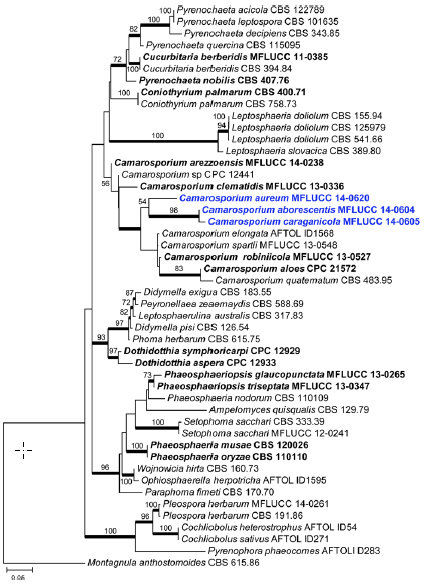
Fig. 1 Phylogram generated from Maximum likelihood (RAxML) analysis based on combined LSU, SSU and ITS sequence data of Pleosporales. Maximum likelihood bootstrap support values greater than 50 % are indicated above or below the nodes, and branches with Bayesian posterior probabilities greater than 0.95 are given in bold. The ex-types (reference strains) are in bold; the new isolates are in blue. The tree is rooted with Montagnula anthostomoides CBS 615.86
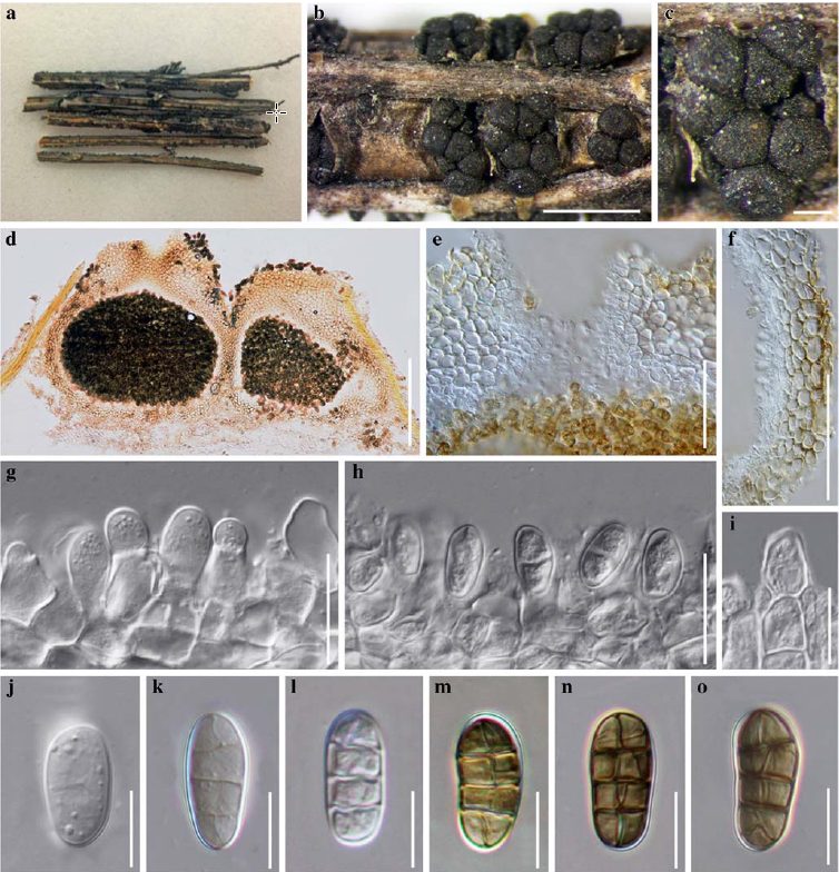
Fig. 2 Camarosporium caraganicola (holotype) a, b Conidiomata on host surface c Gregarious conidiomata on host surface d Vertical section of multi-loculate conidiomata e Ostioles f Part of pycnidium wall g – i Conidiogenous cells and developing conidia j – l Conidia when immature m – o Mature conidia. Scale bars: b = 500 μm, c – e = 200 μm, f = 100 μm, g – h = 20 μm, i – o = 10 μm.