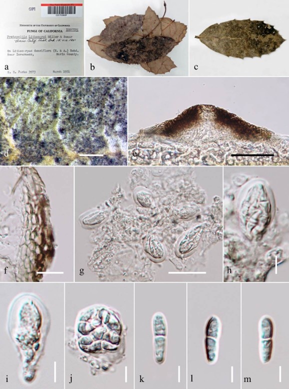
Bonaria lithocarpi (V.A.M. Mill. & Bonar) Bat., Publicações Inst. Micol. Recife 56: 439 (1959) Fig. 22
≡ Protopeltis lithocarpi V.A.M. Mill. & Bonar, University of Calif. Publ. Bot. 19: 412 (1941)
MycoBank number: MB 293763; Index Fungorum number: IF 293763; Facesoffungi number: FoF 05164;
Epifoliar on upper and lower surface of living leaves of Lithocarpus densiflora, forming black rounded spots. Mycelium superficial, not apparently penetrating host cells. Thyriothecia 32– 37 μm high × 148–161 μm diam., circular, solitary or in groups, gregarious, superficial, carbonaceous, dark brown to black, ostiolate. Peridium 6–15 μm wide, comprising a single stratum of black-brown cells of closely arranged textura angularis. Hamathecium comprising asci embedded in pseudoparaphyses. Asci 15–26 μm × 17–25 μm ( x = 21.3–21.4 μm, n = 10), 6-spored, bitunicate, fissitunicate, saccate, globose or subglobose to oblong, apedicellate, apically rounded and endotunica thick-walled near apex without a distinct ocular chamber. Ascospores 14–15 μm × 2–3 μm ( x = 14.7 × 3.3 μm, n = 10), 2 or 4 seriate, elongate-ellipsoidal, hyaline, 2–celled, slightly constricted at the septum, lower cells slightly longer than upper cell, appearing rough-walled. Asexual morph: undetermined.
Material examined – USA, California, Marin, near Inverness, on Lithocarpus densiflora (Fagaceae), March 1931, H.E. Parks. (S-F 3573, isotype).
Notes – Protopeltis lithocarpi was described by Miller & Bonaria (1941). Later, Protopeltis was listed as a synonym of Myriangiella in Schizothyriaceae (von Arx & Müller 1975) and Bonaria was listed as a valid genus in Micropeltidaceae in Lumbsch & Huhndorf (2010).
Economic significance – The genus Bonaria consists of non-obligate parasites which have been reported on stems of Lachnum marginatum (Cooke) Raitv (Momei 2003).

Figure 22 – Bonaria lithocarpi (S-F 3573, isotype) a–c Herbarium specimen and habit on leaf. d Appearance of ascomata on the host surface. e Section of ascoma. f Peridium g Asci embedded in pseudoparaphyses. h–j Asci. k–m Ascospores. Scale bars: d = 500 μm, f = 10 μm, e, g = 20 μm, h– m = 5 μm.
