Austropleospora osteospermi R.G. Shivas & L.Morin,Morin et al., Fungal Diversity 40(1): 70 (2010).
Facesoffungi number: FoF00935; Fig. 1
Parasitic on stem and leaves of Chrysanthemoides monilifera (L.) Norlindh. Sexual morph: Ascomata 75 – 110 × 130 – 200 μm (x̄ = 100 × 146 μm, n = 10), subglobose, sometimes slightly flattened, solitary or in groups, scattered, immersed immediately below the stem epidermis, ostiole 60 – 90 μm long, with a protruding neck. Peridium 8 – 18 μm (x̄ = 12 μm, n = 10) wide, composed of dark brown to black cells of textura angularis. Hamathecium of 2 – 3 μm wide, dense, filamentous, anastomosing, aseptate, hyaline pseudoparaphyses. Asci 75 – 120 × 13 – 18 μm (x̄ = 92 × 16 μm, n = 20), bitunicate, fissitunicate, 6 – 8 – spored, cylindrical to clavate, with a short, broad, pedicel, rounded at the apex, with a minute ocular chamber. Ascospores 16.5 − 21 × 6 – 8.4 μm (x̄ = 18 × 7.7 μm, n = 25), biseriate to overlapping uniseriate, yellowish brown, ellipsoidal, muriform, mostly with 3 – transverse septa, 0 – 2 longitudinal septa, slightly constricted at median septum, not or very slightly constricted at other septa, apex rounded to slightly tapered, base tapered to rounded, smooth. Asexual morph: Conidiomata 75 – 110 × 100 – 130 μm (x̄ = 88 × 115 μm, n = 10), pycnidial, globose, superficial on stem, immersed in the host tissue and becoming erumpent at maturity, globose, dark brown in the erumpent part, with a single ostiole. Conidiomata wall 9 – 16 μm wide, brown to reddish-brown, thin-walled, comprising several layers with cells of textura angularis. Conidiophores reduced to conidiogenous cells. Conidiogenous cells 10 – 12 × 2.5 − 3.5 μm (x̄ = 11 × 2 μm, n = 15), inconspicuously annellidic, discrete, cylindrical. Conidia 14 – 18 × 4.8 − 6.5 μm (x̄ = 15.8 × 5.7 μm, n = 25), cylindrical to narrowly ellipsoidal, initially hyaline and aseptate, becoming yellowish – brown at maturity, mostly transversely 1 – 3 – septate, ends rounded.
Material examined – AUSTRALIA, New South Wales, Bellinger Head, Kalang River mouth, on stem and leaves of Chrysanthemoides monilifera ssp. rotundata (Asteraceae), 5 March 2008, L. Morin (BRIP 52234, holotype).
Notes – Austropleospora was introduced by Morin et al. (2010) to accommodate A. osteospermi and is monotypic. In the same study Shivas and Morin (in Morin et al. 2010) observed Hendersonia osteospermi Wakef. on the same host and identified it as the asexual morph of A. osteospermi both in culture and by DNA sequence analysis. Morin et al. (2010) placed Austropleospora in Pleosporales without assigning it to any family based on ITS sequence analysis. Thambugala et al. (2014) tentatively referred Austropleospora to Pleosporaceae based on morphological similarities but Ariyawansa et al. (2015) excluded Austropleospora from Pleosporaceae and ten tatively referred it to Didymosphaeriaceae.
In the present study we found that the type strain of Austropleospora osteospermi forms a separate clade together with the type strain of Paraconiothyrium archidendri Verkley et al., sister to Paracamarosporium and Paraconiothyrium fuckelii (Sacc.) Verkley & Gruyter species complex clades in the family Didymosphaeriaceae. Thus we assign Austropleospora to the family Didymosphaeriaceae as a distinct genus. Austropleospora forms a distant clade from the generic type of Paraconiothyrium, P. estuarinum and this is also supported by morphology. Paraconiothyrium is paraphyletic within the family Didymosphaeriaceae.
We synonymise Paraconiothyrium archidendri under Austropleospora by giving the priority for the oldest epithet.
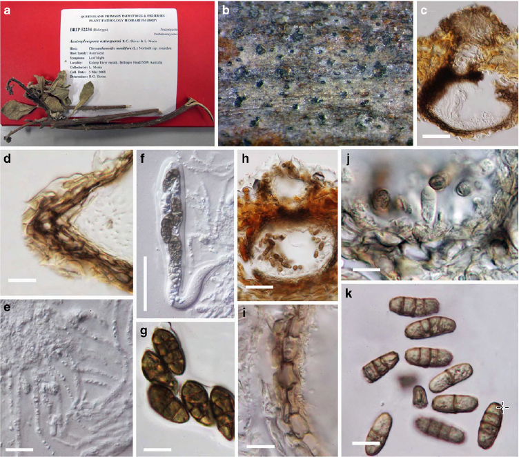
Fig. 1 Austropleospora osteospermi (holotype) a Herbarium material b Ascomata on host surface c Section through ascoma d Section of peridium e Cellular pseudoparaphyses f Mature bitunicate asci g Muriform ascospores h Section of pycnidium i Pycnidial wall j Conidiogenous cells and developing conidia k Brown 3 – septate conidia. Scale bars: c = 50 μm, d = 25 μm, e, g = 10 μm, f = 25 μm, g = 5 μm, h = 20 μm, i – k = 5 μm.