Aulographum hederae Lib., Pl. crypt. Arduenna, fasc. (Liège) 3: no. 272 (1834).
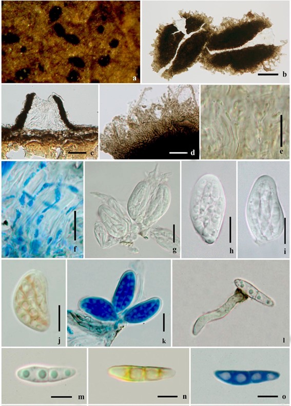
MycoBank number: MB 161393; Index Fungorum number: IF 161393; Facesoffungi number: FoF 07947; Fig. 98
Saprobic on fallen leaves of Hedera helix. Mycelium sparse, not radially arranged. Sexual morph: Ascomata 52-76 μm diam, 35–42 μm high ( x̄ = 72 × 40 μm, n = 5), forming as small black dots on host surfaces, solitary or clustered, nearly superficial, easily removed or breaking from the substrate, globose when immature, becoming sublinear or v-shaped at maturity, rounded, carbonaceous, dark brown to black, with slit-like opening, ostiolar canal filled with hyaline cells, with cells comprising parallel, radiating lines from center of the ostiole to outer cells, becoming pale brown at the margin. Hamathecium comprising 1 µm wide, septate, anastomosing, branched, trabeculate pseudoparaphyses, embedded in a gelatinous matrix with asci. Asci 24–30 × 10–15 μm ( x̄ = 28–12 μm, n = 10) 8-spored, bitunicate, fissitunicate, oval to ellipsoid, apex thickened, short pedicellate or lacking, and with a wide, indistinct ocular chamber. Ascospores 11–14 × 3–5 μm ( x̄ = 12–3 μm, n = 10) overlapping 2–3-seriate, oblong to ovate, narrow at both ends, hyaline, 1-septate at the center, constricted at the septum, the upper cell often broader than lower cell, smooth-walled. Asexual morph: Undertermined.
Material examined: Germany, Frankfurt, on fallen leaves of Hedera helix, in 2012, Meike Piepenbring MFLU, culture MFLUCC 12-0397 (MFU) = CPC21373 (CBS).
Notes: Aulographum hederae is the type species of Aulographaceae. It is characterized by longitudinally splitting ascomata and a perithecial wall appressed by mycelium with bright coloured margin (Hongsanan et al. 2014b). Sequence data of this specimen was provided by Hongsanan et al. (2014b), but without a plate and description. There- fore, we provide a photoplate of the same specimen with descriptions.

Fig. 98 Aulographum hederae (MFLUCC 12-0397). a Habit, ascomata on host substrate. b Ascomata with slit-like opening. c Section through ascoma. d Walled cells of ascoma. e, f Hamathecium in Melzer’s reagent and Cotton blue reagent respectively. g–i Asci. j Asci in Melzer’s reagent. k Asci in Cotton blue reagent. l Ascospore germinated. m–o Ascospore in water, Melzer’s reagent and Cotton blue reagent respectively. Scale Bars: b = 50 µm, c, g–k = 20 µm, d–f = 10 µm, l–o = 5 µm
