Asperosporus subterraneus, Karl.-Ayala, Gazis & M.E. Sm., sp. nov.
MycoBank number: MB 838906; Index Fungorum number: IF 838906; Facesoffungi number: FoF; Figs 1, 3A–F.
Etymology: From the Latin “sub” (below) and “terra” (earth), for the habit of fruiting below the soil
Diagnosis: Basidiomata 5–30 mm diam, globose to irregular, astipitate, light tan but drying dark brown and staining pink-red when handled, producing a rancid odor when fresh. Peridium thin and friable when dry. Gleba loculate, staining pink-red when exposed to air, friable and hydrophobic when dried. Basidia 1–3 spored in fresh specimens, collapsing upon drying. Spores 16–22 × 12–18 μm, subglobose or ellipsoid, ornamented with warts, thick walled, strongly dextrinoid, often with sterigma remnants remaining attached. The genus is presently monotypic.
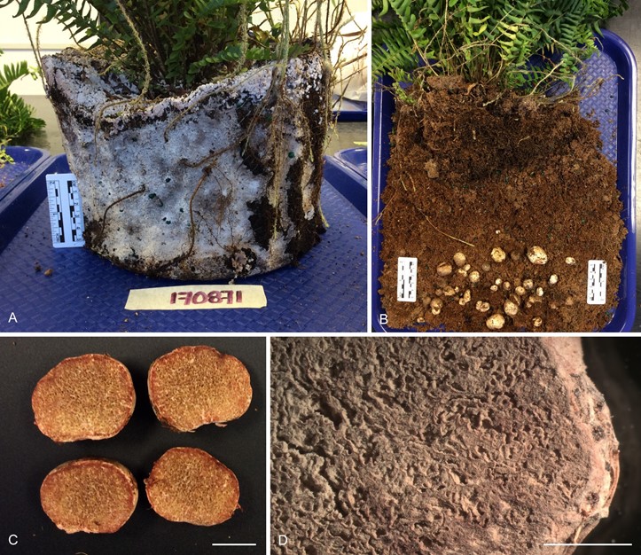
Macroscopic features: Basidiomata 5–30 mm diam, globose to irregular, lacking a stipe, surface smooth, at first white or light tan but becoming brownish with age, staining pink-red where handled or bruised (Fig. 1C), drying darker brown, particularly where bruised. Peridium irregular, friable when dry (Fig. 1D), sometimes sloughing off completely where handled, staining dark brown to black in Melzer’s reagent. No response in KOH when dried. Gleba compressed, irregular, loculate (Fig. 3A), lacking a columella, light tan at first with occasional veins of white trama tissue and pockets of darker brown spores, staining pinkish or red when cut and exposed to air, reddish staining more notable in younger specimens, turning dark brown, powdery, delicate and hydrophobic upon drying. Odor rancid when fresh, indistinctly fungal when dry. Taste not determined.
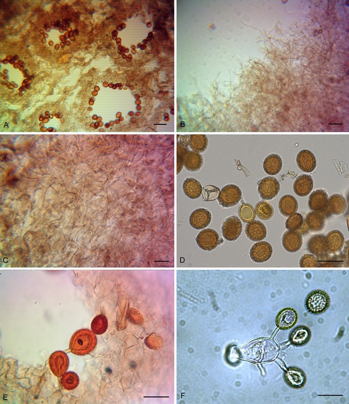
Microscopic features: Basidiospores 16–22 × 12–18 μm (av. 18 × 15 μm) at maturity, globose, subglobose or ellipsoid (Q = 1–1.47 μm, mean Q = 1.21 μm); walls 1–3.5 μm thick (av. =1.8 μm); restricted at the sterigmal attachment point and apiculate, notably ornamented with larger pyramidal to irregular warts, up to 1 μm tall and 1 μm wide. Immature spores are notably smaller, mostly 12–15 × 10–12 μm, and with smaller spines that are 0.5 × 0.5 μm and clearly separated from one another.
Sterigma remnants often remaining attached and clearly visible in many spores (Fig. 3E) but more common in young spores, most 4–5 μm × 1–2 μm but sometimes up to 9 μm long. Pale yellow- orange when young but becoming darker orange-brown at maturity when observed using KOH or water, strongly dextrinoid in Melzer’s reagent with mature spores turning notably darker than younger spores, highly variable in size and shape. Basidia 1–3 spored, difficult to find and see, collapsing in mature dried specimens (Fig. 3F). Cystidia not observed. Peridium (100–)150– 250(–350) μm thick, composed of loosely interwoven (Fig. 3B) and irregularly branched and septate, single layered, hyphae 3–5 μm diam, occasionally swelling up to 10 μm; arrangement of hyphae mostly tangled and irregular but occasional bands of hyphae parallel to the exterior near the peridial surface, light yellow-brown, some hyphae strongly dextrinoid. Trama tissue 75–200 μm thick, composed of irregularly shaped, elongated and inflated hyaline hyphae, 10–26 μm diam. Subhymenium approximately 10–40 μm thick, comprised of densely packed interwoven hyphae with cells 12–14 μm diam that are brown to orange-brown. Clamp connections absent on all hyphae. No response to KOH.
Habitat and distribution: Found in south Florida growing in soil of potted nursery plants with poor drainage. Specimens thus far found only in association with Boston Fern (N. exaltata) which was planted in a Canadian peat moss and Florida pine bark mixed potting soil during the winter months.
Typus: USA, Florida, Tropical Research and Education Center Plant Diagnostic Clinic, Homestead, Miami-Dade Co., 21 Dec. 2017, R. Gazis, MES-3094 (holotype FLAS-F-68001).
Additional collection examined: USA, Florida, Tropical Research and Education Center Diagnostic Clinic, Homestead, Miami-Dade Co., 4 Dec. 2018, R. Gazis, 180871 (Photos deposited in MycoBank, but specimens not dried properly and therefore discarded).

Fig. 1. Asperosporus subterraneus specimen FLAS-F-68001 (holotype) A. White hydrophobic mycelial mat binding organic matter in one of the potted Boston Fern plants (Nephrolepsis exaltata) submitted to the University of Florida Plant Diagnostic Clinic. B. Gasteroid basidiomata found in the potting soil. C. Red-staining gleba when freshly cut. D. Dry powdery gleba in a dried specimen, showing locules and the thin friable peridium. Scale bars: C–D = 1 cm.

Fig. 3. Microscopic features of Asperosporus subterraneus specimen FLAS-F-68001 (holotype) A. Loculate gleba B. Peridium hyphae. C. Tangled peridium hyphae. D. Ornamented spores with thick spore walls. E. Subhymenium with basidiospores, some of which have retained sterigma remnants (indicated with arrows). F. Basidium with basidiospores. Scale bars: A–C = 60 μm, D–F = 20 μm.
