Ascochyta medicaginicola var. medicaginicola Q. Chen & L. Cai 2015
≡ Phoma medicaginis var. medicaginis Malbr. & Roum., Rev. Mycol. 8: 91. 1886.
MycoBank number: MB 623214; Index Fungorum number: IF 623214; Facesoffungi number: FoF 10670;
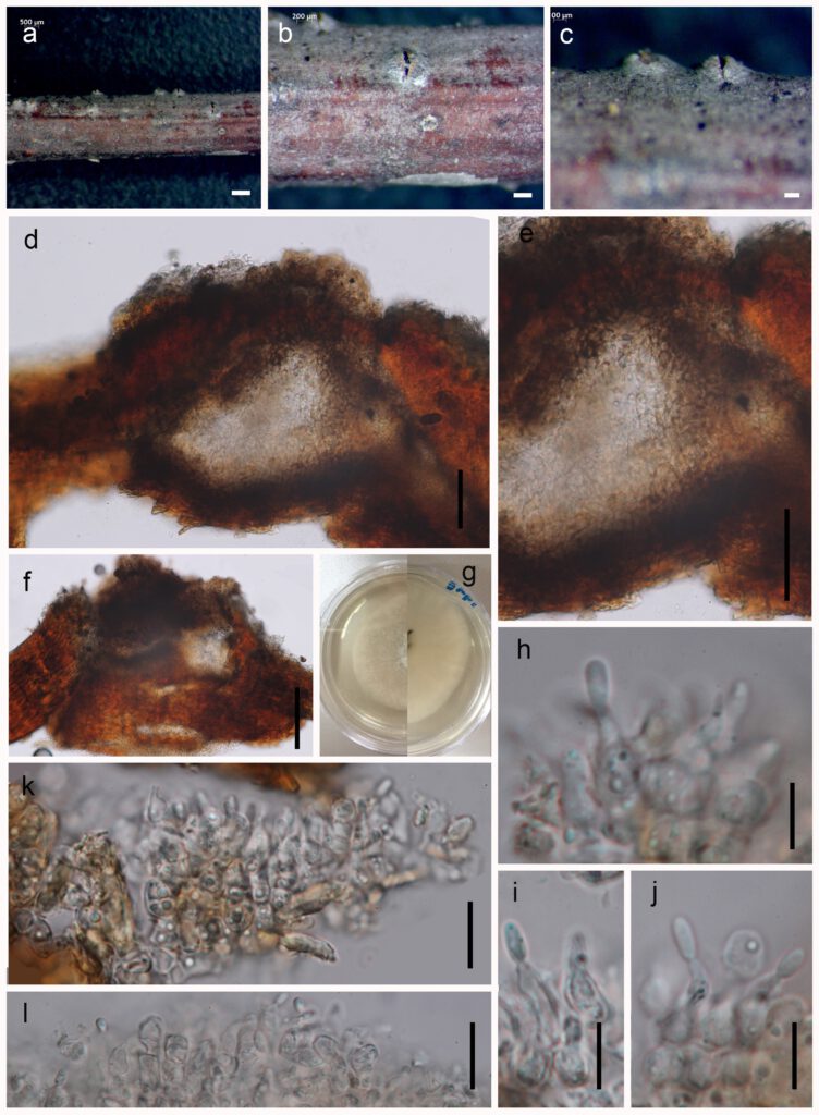
Pathogenic on living branches of Prunus cerasifera noticeable as black, circular dots on the host surface. Asexual morph: Conidiomata 165–190 μm high, 170–210 μm wide, black, scattered or gregarious, superficial to immerse on the substrate, black, subglobose to globose, uniloculate. Ostiolar neck 25–50 lm long, 3–5 lm wide, covered with 1-celled, thick-walled, dark brown to almost black. Peridium 15–25 lm wide at the base, 30–80 lm wide at the sides, thick, comprising 3–4 layers, outer most layer heavily pigmented, thick-walled, comprising blackish to dark brown loosely packed cells of textura angularis, inner layer composed 3–5 layers, pale brown to hyaline, cells towards the inside lighter, flattened, thick-walled cells of textura angularis. Sexual morph: Not observed.
Material examined: RUSSIA, Rostov region, Shakhty, near railroad, on dead twigs of wild tree of Prunus cerasifera Ehrh. (Rosaceae), 11 May 2017, Timur S. Bulgakov, T-1832 (MFLU 17-2138, holotype); ex-type living cultures, MFLUCC 18–0453. GenBank ITS: ***; LSU: ****.
Notes: This is the record of Ascochyta medicaginicola and characters clearly indicate that it is identical to A. medicaginicola var. medicaginicola (Boerema et al. 2004, Chen et al. 2015). The multigene phylogenetic analysis shows that this collection groups in the Ascochyta medicaginicola clade with 52 MLBS support (Fig. 1). This collection also fits with the generic concept of Ascochyta in morpholohy of asexual morph of Ascochyta medicaginicola and the phylogenetic analysis show the topology same with Jayasiri et al. 2017.

Figure captions:
Ascochyta medicaginicola var. medicaginicola a, b, c Conidia observed on host substrate. d, e Conidiomata. f, g, h Conidia i, k, l Conidia j Conidiodenous cell. Scale bars: a = 500 µm, b, c, d, e, g = 100 µm, f,h = 50 µm, i, j, k, l = 10 µm.
