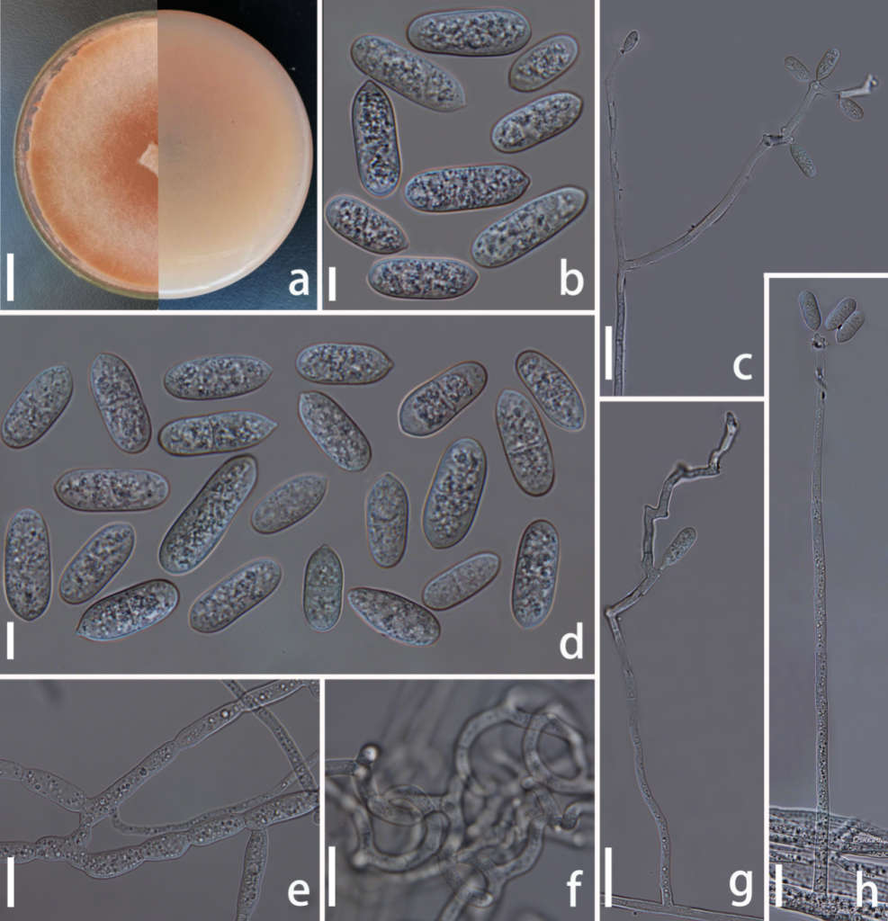
Arthrobotrys shuifuensis F. Zhang, X.Y. Yang & K. D. Hyde, sp. nov. FIGURE 5.
MycoBank number: MB; Index Fungorum number: IF; Facesoffungi number: FoF 10764;
Etymology: The species name “shuifuensis” refers to the name of sample collection site: Shuifu County, Zhaotong City, Yunnan Province, China.
Description: Colonies on PDA initially white and turned to tinged pink after two weeks, cottony, rapidly growing, reaching a diameter of 50 mm after 9 days incubation at 26°C. Mycelium colourless, composed of septate, branched, smooth hyphae. Conidiophores hyaline, erect, septate, unbranched or rarely branched, producing several separate nodes by repeated elongation of conidiophores, with each nodes consisting of 2–8 papilliform bulges and bearing polyblastic conidia, 105–305 µm (x̅ = 218.2 µm, n = 50) long, 3–5 µm (x̅ = 3.8 µm, n = 50) wide at base, gradually tapering upwards to a width of 1.5–3.5 µm (x̅ = 2.5 µm, n = 50) at the apex. Conidia hyaline, smooth, capsule-shaped, with one median septum, 17–36 × 5–12.5 µm (x̅ = 27.2 × 8.2 μm, n = 50). Chlamydospores hyaline, smooth, cylindrical, in chains, 6–18 × 3–7.5µm (x̅ = 9.7 × 8.2 μm, n = 50). Capturing nematodes with adhesive network.
Material examined: CHINA, Yunnan Province, Zhaotong City, Shuifu county, from terrestrial soil, 16 June 2017, F. Zhang. (CGMCC3.19716, Holotype!).
Sequence data: ITS:MT612334.1

FIGURE 5. Arthrobotrys shuifuensis (CGMCC3.19716, holotype!). (a) Colony. (b, d) conidia. (e) Chlamydospore. (c, g, h) Conidiophore. (f) Trapping-device: adhesive network. Scale bars: (a) = 1cm, (b, d, e) = 10 µm, (c, f–h) = 20 µm.
