Aquihelicascus thalassioideus (K.D. Hyde & Aptroot) W. Dong & H. Zhang, comb. nov.
MycoBank number: MB 557920; Index Fungorum number: IF 557920; Facesoffungi number: FoF09262; Fig. 79
Basionym: Massarina thalassioidea K.D. Hyde & Aptroot, Nova Hedwigia 66(3–4): 498 (1998)
Synonymy: Helicascus thalassioideus (K.D. Hyde & Aptroot) H. Zhang & K.D. Hyde, Sydowia 65(1): 159 (2013)
Freshwater distribution: Australia (Hyde and Aptroot 1998b), Brunei (Hyde and Aptroot 1998b), China (Tsui et al. 2000; Ho et al. 2001; Luo et al. 2004), French West Indies, Martinique (Zhang et al. 2015), Peru (Shearer et al. 2015), Janpan (Tanaka et al. 2015), Philippines (Hyde and Aptroot 1998b), Thailand (Kurniawati et al. 2010; Zhang et al. 2013a; this study)
Saprobic on decaying wood submerged in freshwater. Sexual morph: Pseudostromata clustered, immersed, visible on the host surface as blackened ostiolar dots. Pseudoparaphyses 1–3 μm diam., abundant, cellular, hypha- like, hyaline, septate, embedded in a gelatinous matrix. Asci 150–190 × 14.5–23 μm (x̄ = 175 × 17.5 μm, n = 5), 8-spored, bitunicate, fissitunicate, clavate, long pedicellate, up to 90 μm long. Ascospores 24–28 × 7–10 μm (x̄ = 25.5 × 8.5 μm, n = 25), overlapping uni- to bi-seriate, some- times overlapping tri-seriate, ellipsoidal, hyaline, 1-septate, constricted at septum, curved, thin-walled, smooth. Asexual morph: Undetermined (detailed description see Hyde and Aptroot (1998b) and Zhang et al. (2013a)).
Material examined: THAILAND, Chiang Rai Province, on submerged wood in a stream, 1 July 2018, W. Dong, CR134-1 (MFLU 18-1705), living culture KUMCC 19-0094; ibid., CR134-2 (HKAS 105076).
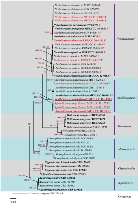
Notes: Our new collection KUMCC 19-0094 is identified as Aquihelicascus thalassioideus based on multigene phylogenetic analysis (Fig. 84). The morphological features of our collection also fits well with A. thalassioideus except for the long pedicel, which is probably because of the pedicel spreading in water (Fig. 79). Most publications do not report an ascospore sheath for A. thalassioideus except Zhang et al. (2015) who noted some fugacious mucilaginous remnants visible in Indian Ink when ascospores were just released from the asci. This character was not observed in our collection.

Fig. 79 Aquihelicascus thalassioideus (MFLU 18-1705). a, b Immersed pseudostromata with blackened ostiolar dots. c Ascus embedded in pseudoparaphyses. d, e Bitunicate asci. f, g Ascospores. Scale bars: c–e = 50 μm, f, g = 20 μm

Fig. 84 Phylogram generated from maximum likelihood analysis of combined LSU, SSU, ITS and TEF sequence data for species of Morosphaeriaceae. Bootstrap values for maximum likelihood equal to or greater than 75% and Bayesian posterior probabilities equal to or greater than 0.95 are placed near the branches as ML/BYPP. Newly generated sequences are in red and ex-type strains are in bold. The new species introduced in this study are indicated with underline. Freshwater strains are indicated with a red letter “F”. The tree is rooted to Latorua caligans CBS 576.65 (Latoruaceae)
