Apoharknessia insueta (B. Sutton) Crous & S.J. Lee, Stud. Mycol. 50: 240. (2004).
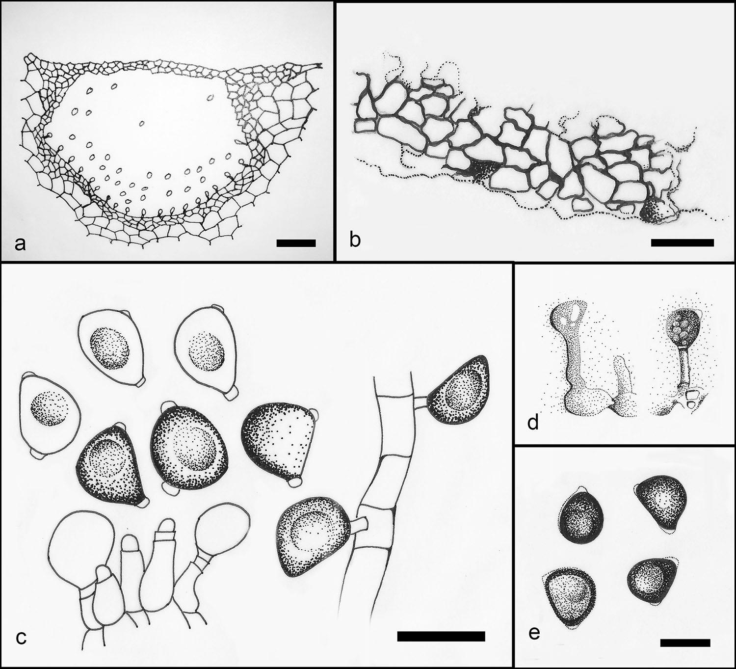
Foliicolous forming bleached spots or saprobic on various substrates. Sexual morph: Undetermined. Asexual morph: Conidiomata stromatic, subepidermal to immersed, solitary to gregarious, subglobose to irregular, unilocular, pale brown. Conidiomata wall outer layer composed of thin-walled, pale brown cells of textura angularis, inner layer pale yellow to hyaline. Conidiophores reduced to conidiogenous cells. Conidiogenous cells 5–15 × 4–6 µm (x̅ = 9 × 4.8 µm), lageniform to ampulliform, hyaline, smooth, invested in mucus. Conidia 10–12 × 7.5–9 µm (x̅ = 10.5 × 8 µm), conical, aseptate, brown, with a longitudinal band on the flat surface, thick and smooth-walled, guttulate, with short hyaline apiculus, with small globule of mucus on base. Basal appendage 2 9 1–1.5 µm, often gelatinizing and resulting in a minute marginal frill on the truncate base of the conidia (description based on Nag Raj 1993).
Notes: Apoharknessia was introduced and typified by Harknessia insueta and this genus currently comprises two
species. Morphologically and phylogenetically Harknessia shows close affinity to Lasmenia (Senanayake et al. 2017b).
Fig. 1 Consensus tree resulting from a RAxML analysis of a combined nrITS and nrLSU, sequence alignment for taxa of Apoharknessiaceae. Genera are indicated in coloured blocks. Bayesian posterior probabilities are given at the nodes. The scale bar represents the expected number of changes per site.

Fig. 2 Apoharknessia insueta (redrawn from Lee et al. 2004). a Cross section of conidiomata. b Peridium. c–d Conidiogenous cells attached to conidia. e Conidia. Scale bars: a = 50 µm, b–e = 10 µm.