Apiosporopsis carpinea (Fr.) Mariani, Atti Soc. ital. Sci. nat. (Modena) 50: 165. 1911.
Basionym: Xyloma carpini Fr., Observ. mycol. (Havniae) 2: 363. 1818.
Synonymy: See Index Fungorum (2018)
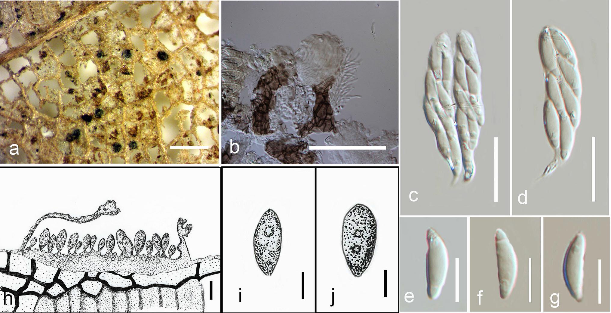
Saprobic on over-wintered plants. Sexual morph: Clypeus 70–140 µm wide, 50–70 µm high, slight, prosenchymatous. Ascomata 112–250 µm diam., 140–170 µm high, globose or depressed, immersed, usually hypophyllous, apapillate, apex rounded with plane pore or short papillate or conic. Peridium 10–20 µm wide, comprising thickwalled, brown cells of textura angularis. Asci 40–75 × 8–14 µm (x̅ = 55 × 10 µm, n = 15), 8-spored, unitunicate, cylindrical, sessile, apical ring bilobed, distinct, shallow. Ascospores 10–15 × 3.5–6.5 µm (x̅ = 11 × 5 µm,
n = 15), overlapping uniseriate, ellipsoid, ovoid or fusoid, straight or often inequilateral, guttulate, hyaline, aseptate.
Asexual morph: Conidiomata acervular, superficial, black, coriaceous. Conidiophores reduced to conidiogenous cells. Conidiogenous cells 5–10 µm long, conical, wide, aseptate, hyaline. Conidia 12–15 × 8–9 µm, oblong to ellipsoid, hyaline, aseptate, with two small guttules (description based on Senanayake et al. 2017b).
Material examined: AUSTRIA, Sonntagberg, New Rosenau, July, on leaves of Carpinus betulus L. (Betulaceae),
P.P. Strasser, IMI 11662.
Notes: Apiosporopsis was introduced based on Apiosporopsis carpinea (= Xyloma carpini Fr.), however Mariani (1911) designated A. saccardiana as the type species. We confirm Apiosporopsis carpinea as the type species, considering the availability of molecular data and good morphological descriptions. Apiosporopsis carpinea was recorded only on over-wintered living leaves. Gloeosporium robergei was reported as the asexual morph of A. carpinea (Potebnia 1910; Treigien and Markovskaja 2007), but there is no molecular data to prove this. Gloeosporium robergei was reported as the causal agent of bud mortality and twig cankers on Ostrya virginiana (Sinclair and Hudler 1980).
Fig. Apiosporopsis carpinea (IMI 11662, redrawn from Potebnia 1910). a Ascomata on substrate. b Vertical section of ascomata. c–d Asci. e–g Ascospores. h Cross section of conidioma. i –j Conidia. Scale bars: a = 500 µm, b = 50 µm, c–g = 10 µm, h = 20 µm, i–j = 5 µm.