Alloscorias syngonii Haituk & Cheew., in Haituk, Suwannarach, Hongsanan, Senwanna & Cheewangkoon, Phytotaxa 507(4): 276 (2021)
Index Fungorum number: IF 558108; FacesofFungi number: FoF 11069
Etymology — Refers to the host, on which the sexual morph of the taxon was collected.
Ex-type culture — SDBR-CMU355
Epiphytic on the upper surface of Syngonium podophyllum leaves. Thallus very thin, superficial, pale brown to brown walls. Sexual morph: Ascomata 47–66 μm diam., 38–64 μm high, (x̅ = 54.9 × 54.4 μm, n = 20), gregarious to scattered, superficial, globose to subglobose, brown to dark brown, with indistinct ostiole. Peridium 20–24 μm. wide (x̅ = 21 μm, n = 10), composed of two sections, arranged in textura angularis, outer layer of brown to dark brown, thick-walled cells, inner layer of light brown thin-walled cells. Setae 30–41 μm long × 3–4 μm wide (x̅= 36.1 × 3.7 μm, n = 20) abundant, corniform, brown, 4–7-septate, constricted at septa, smooth-walled. Asci 27–33 × 14–16 μm (x̅ = 29.7 × 15.4 μm, n = 10), 8-spored, bitunicate, obpyriform sessile, rounded at the apex. Ascospore 14–17 × 2–5 μm (x̅ = 16 × 3.6 μm, n = 10), overlapping 2–3-seriate, hyaline, smooth-walled, fusoid, narrowly rounded at the ends, 3–4-septate, upper cells wider and shorter than lower cells, constricted at the septa, with a conspicuous mucilaginous sheath. Asexual morph: Mycelium pale brown to brown at the apex, septate, constricted at the septa, rough, thick-walled. Coelomycetous: Conidiomata 42–92 μm high × 32–76 μm diam. (x̅ = 73 × 49 μm, n = 10), solitary, brown to dark brown, globose to subglobose. Pycnidial walls composed of pseudoparenchymatous cells of textura angularis, thick-walled, outer layer brown to dark brown, inner layer with hyaline cells. Conidiophores reduced to conidiogenous cells. Conidiogenous cells enteroblastic, phialidic, integrated, terminal, smooth, hyaline, thin-walled. Conidia 4.7–7.4 × 1.7–2.6 μm (x̅ = 5.8 × 2.2 μm, n = 30), oblong to ellipsoid, hyaline, aseptate, guttulate, smooth-walled.
Culture characteristics — Ascospores germinated on PDA within 20–24 hr at 25−30°C, colonies on PDA reaching 2.5–5 mm diam. after one month, greenish brown, leathery, circular, dense, aerial, reverse dark brown, filamentous. Mycelium, brown, pale brown at the apex, septate, constricted at the septa, rough and thick-walled, with a mucilaginous outer wall layer. Colonies on OA reaching 6–10 mm diam, producing dense, irregular, flat or effuse, slightly raised, olivaceous grey. Asexual morph sporulated after two months on OA at 25°C, visible as water drop-like on colony.
Materials examined — Thailand, Mae Hong Son Province, Mueang District, Mok Champae Sub-district, on living leave of Syngonium podophyllum (Araceae), 16 September 2019, Sukanya Haituk, Culture number SDBR-CMU355 (Holotype and Ex-type), TBRC 14598.
Notes — Based on morphological comparison with the type species of other genera in Readerielliopsidaceae, A. syngonii is similar to Scorias spongiosa (MFLUCC 10-0084) in having ascomata globose to subglobose, brown to dark brown and hyaline ascospore (Hughes 1976, Chomnunti et al. 2011). However, A. syngonii have thallus setae and ascospore with a mucilaginous sheath. The asexual morph of A. syngonii produced in globose conidiomata, while Scorias spongiosa produces in flask-shape conidiomata. In addition, Alloscorias is similar to Trichopeltis (Trichopeltinaceae), especially Trichopeltina asiatica, which also appears “root”-like, with ascospores having only transverse septa (Hongsanan et al. 2015b). However, it differs in having mucilaginous sheath ascospore. In a BLASTn search on NCBI GenBank, the closest matches of LSU sequence data of A. syngonii are S. leucadendri with 98% identity to the strain CBS 131318, while the closest matches with the ITS sequence was with 82% S. spongiosa strain CBS 325.33.
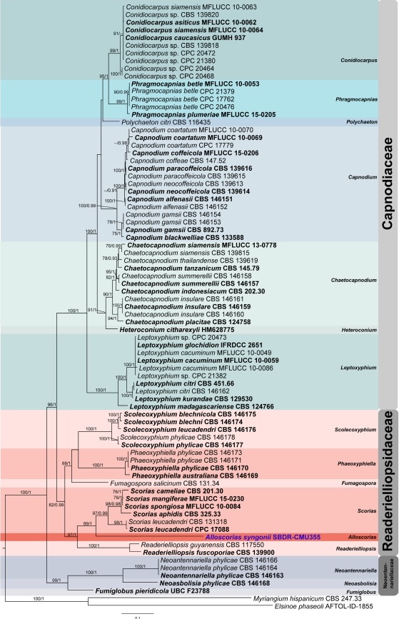
FIGURE 1. Phylogram generated from the best scoring of the RAxML tree based on a combined LSU, ITS, TEF-1α and RPB2. Bootstrap support values for maximum likelihood higher than 75% and Bayesian posterior probabilities (BYPP) greater than 0.90 are displayed above the nodes, respectively (ML/BYPP). The ex-type strains are in bold. Newly generated sequences are indicated in blue. The tree is rooted to Myriangium hispanicum (CBS 247.33) and Elsinoe phaseoli (AFTOL-ID-1855).
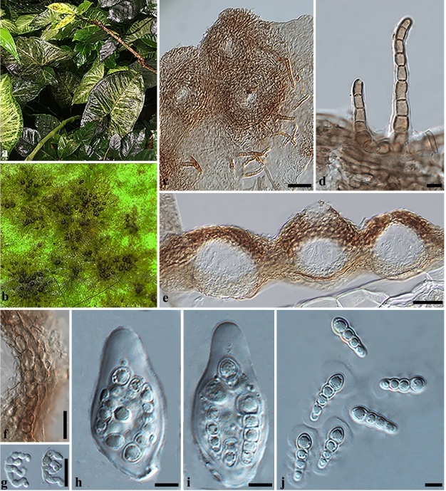
FIGURE 2. Sexual morph of Alloscorias syngonii (SDBR-CMU355, holotype). a Epiphytic sooty mold on host. b Ascomata on surface of leaves. c Thallus and setae on host substrate. d Setae. e Section of ascomata. f Section through peridium. g Germinating ascospore. h–i Asci. j Ascospores with mucilaginous sheath. Scale bars: c, e = 30 µm; f–g = 20 µm; d, h–j = 5 µm.
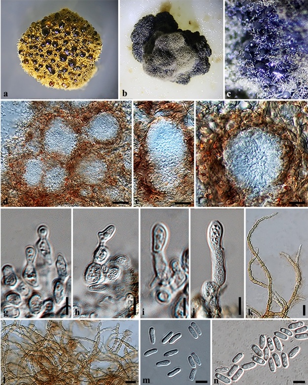
FIGURE 3. Asexual morph of Alloscorias syngonii (ex-type culture). a Culture characteristics on PDA (3-wk-old). b Culture characteristics on OA (3-wk-old). c Colonies sporulating on OA (3-wk-old). d–f Section of conidioma. g–i Conidiogenous cells and developing conidia. k–l Mycelium. m Conidia in lactic acid. n conidia in Indian ink revealing guttulate. Scale bars: d = 30 µm, e–f, l = 20 µm, k = 10 µm, g–j, m–n = 5 µm.