Atrozythia klamathica J.K. Mitch. & Quijada, in Mitchell, Garrido-Benavent, Quijada & Pfister, IMA Fungus 12 (no. 6): 17 (2021)
Index Fungorum number: IF 838700; MycoBank number: MB 838700; Facesoffungi number: FoF 15028;
Etymology – Named after the collection locality of the holotype, Klamath National Forest.
Type – USA: California: Siskiyou County, Klamath National Forest, southwest side of Forest Route 17N11, 41°50’03.6″ N 123°25’42.1” W, 566 m a.s.l., apothecia on resinous wounds of living young Chamaecyparis lawsoniana, 12 Dec. 2017, J.K. Mitchell JM0068 (FH 00965406 – holotype).
Sexual morph: Apothecia discoid to cupulate, scattered, erumpent from the resin, consistency coriaceous and ascomata slightly shrunken when dry, but expanding and fleshy when moist, 0.7–1.2 mm diam, to 1 mm high, subsessile to short-stipitate (0.1–0.3 × 0.2–0.3 mm), stipe narrower toward the base. Disc concave to plane, round or somewhat irregular by internal growing pressure, smooth or slightly wrinkled, black (267.black) to dark greyish brown (62.d.gy. Br), with or without light white (263) to light blue grey (190) coating pruina; margin distinct, raised when immature but not protruding beyond the hymenium when mature, 0.5–1 mm thick, entire and smooth or radially cracked, concolourous with hymenium and usually pruinose. Receptacle concolourous with hymenium and margin, strongly roughened, more heavily pruinose, pruina extending downward on the stipe, anchoring hyphae surrounding the receptacle from the base of the stipe to the lower flank and rarely at the margin; pruina can be lost during development and is usually more frequent in immature apothecia. Asci (103–)131–158(–166) × (27.5–)29.5–36.5(–40.5) μm, cylindric-clavate, multi-spored, mature asci 35–50 μm below the hymenial surface prior to spore discharge, ascus dehiscence rostrate, inner wall material expanding, protruding c. 40–50 μm, reaching the hymenial surface at spore discharge; apex hemispherical, thick-walled, strongly staining in CR, apex with an apical chamber, apical wall 3–5 μm thick, chamber later disappearing and apical tip thickening, be- coming 10–15.5 μm thick, projecting into the ascus, be- coming dome-like, with intermediate morphologies also observed, inner wall not or faintly amyloid, outer wall in- tensely amyloid; lateral walls 1–3.5 μm thick, asci covered with an amyloid gel layer; base arising from a perforated crozier. Ascospores 1.8–2.3 μm diam, globose to subglobose, hyaline, inamyloid, aseptate, wall slightly thick, and with one eccentric medium grey (265) lipid guttules. Paraphyses embedded in a thick, hyaline layer of gel, cylindrical, uninflated to medium clavate, straight or slightly wavy, terminal cell (5.5–)6.5– 9(–11.5) × 2–3.3(–4.5) μm, covered by a strong yellowish brown (74) to deep yellowish brown (78) amorphous exudate, lower cells (6.5–)8.5–11 × 2–3 μm, basal cells (12.5–)14.5–18(–20.5) × 1.5–2 μm, simple, unbranched, hyaline, septate, septa strongly staining in CR, basal cells ± equidistantly septate, terminal and lower cells shorter, walls smooth, sparse tiny yellow grey (93) lipid guttules throughout, from the basal to terminal cells. Excipulum composed of two differentiated layers sharply delimited, ectal excipulum strongly gelatinized, (111–)127–165(–192) μm thick at lower flank and base, (32–)48–124(–132) μm thick at margin and upper flank, constituted of three layers; innermost layer of moderately packed textura intricata immersed in a pigmented gel, strong brown (55) to dark brown (59), with sparse dark greyish yellow (91) refractive amorphous lumps; middle layer with loosely packed hyaline cells, strongly gelatinized, parallel to each other (sometimes interwoven) and oriented perpendicular to the outer surface, outermost layer with shorter, parallel and very tightly packed cells without intercellular spaces, walls pigmented and surrounded by a strong brown (55) to dark brown (59) amorphous exudate, cortical layer irregular and black (267). Individual cells at the middle layer of the ectal excipulum (5–)6.5– 9(–10) × 2–3.5 μm at the margin, (6.5–)8.5–12(–15.5) × 2–3 μm at the lower flank and base cell walls 0.5–1.5(–3.5) μm thick. Medullary excipulum of slightly gelatinized textura intricata, tightly packed, cells neither with intercellular spaces nor particular orientation, (10–)12.5–16.5(– 19) × 2–3(–3.5) μm. Asexual morph: unknown.
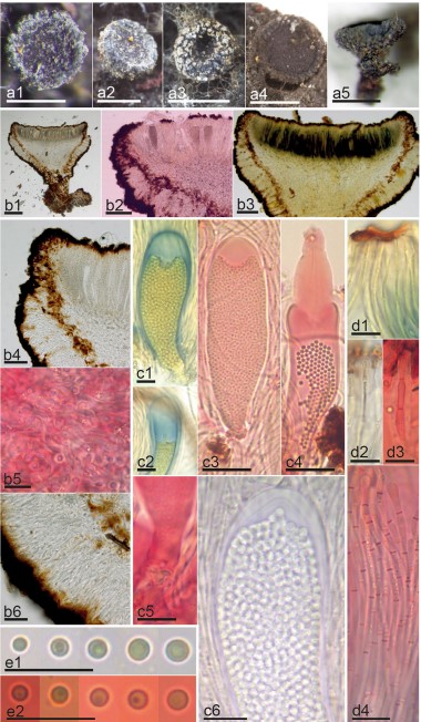
Notes – This species is known from two specimens (of which the holotype was sequenced twice) and is illustrated in Fig. 3. It was probably also observed once in Alaska (Goff 2020), but no specimen was collected. Little is known about its ecology or possible asexual morphs. Sequence and morphological data are sufficient to separate it from Sarea and Zythia, and it shows a closer affinity to the latter. Although apparently collected only twice, it is possible (given the rarity with which Sarea difformis is found on cupressaceous hosts) that A. klamathica is the fungus which was isolated as an endophyte of cupressaceous plants in central Oregon and reported as S. difformis (Petrini and Carroll 1981) but due to the lack of detailed data in the report, that support can neither be confirmed nor refuted. Culture work with fresh material should be done.

Fig. 3 Morphological features of Atrozythia klamathica. a1–5 Dry apothecia. b1–6 Median section of apothecium with details of excipulum: b4. Ectal excipulum at margin, b5. Medullary excipulum, b6. Ectal excipulum at lower flanks. c1–6 Morphological variation of asci: c1–2. Amyloid walls, c3. multispored mature ascus, c4. Ascus dehiscence, c5. Perforated crozier, c6. Details of ascus walls. d1–4 Paraphyses. e1–2 Ascospores. Reagents: H2O = b4, b6; KOH = b1, c6, e1; KOH+CR = b2, b5, c3–5, d1, d3–4, e2; KOH+MLZ = b3, c1–2, d2. Scale bars: 500 μm = a1–5; 200 μm = b1; 100 μm = b2–3; 50 μm = b4, b6, c3–4; 10 μm = b5, c1–2, c5–6, d1–4, e1–2. Collections: JM0068 = a1–2, a5, b1–6, c5–6, d2–4, e1; Haldeman 2748 = a3–4, c1–4, d1, e2
