Pseudocamarosporium propinquum (Sacc.) Wijayaw., Camporesi & K.D. Hyde, in Wijayawardene, et al. Cryptog. Mycol. 35(2): 191 (2014)
Index Fungorum Number: IF550560; Facesoffungi number: FoF 00020
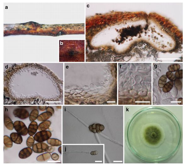
Saprobic on Tamarix gallica L. Sexual morph: Undetermined. Asexual morph: Conidiomata pycnidial, 200–400 μm diam. × 120–200 μm high (x = 323 × 168 μm, n = 6), solitary, scattered, black, immersed, unilocular, subglobose or irregular in shape, frequently associated with other fungi, ostiolate. Pycnidial wall 14–25 μm (x = 18.9 μm, n = 12), comprising 3–4 layers of hyaline to light brown cells of textura angularis and indistinguishable from the host tissues. Conidiophores reduced to conidiogenous cells. Conidiogenous cells 4–6×5–8 μm (x = 12.6 × 7 μm, n = 30), with percurrent phialidic development, smooth, short, hyaline, formed from the inner layer of the pycnidial wall. Conidia 10–15 × 5.5–8 μm (x = 12.6 × 6.5 μm, n = 30), oblong, straight, initially hyaline, aseptate, becoming muriform, with 3 transverse septa and 1–2 longitudinal septa, pale brown to dark brown, smooth-walled.
Culture characteristics: Conidia germinating on PDA within 24 h and germ tubes produced from one or several septa. Colonies growing on PDA reaching 47 mm diam. After 14 days at 25 °C, circular, flat, moderately dense, surface greenish olivaceous, white at the margin, becoming dark olive to greyish after 2 weeks, reverse black, smooth surface with entire to slightly undulate edge.
Material examined: ITALY, Province of Forlì-Cesena, Ravaldino in Monte – Forlì, Tamarix gallica L. (Tamaricaceae), 10 January 2014, Erio Camporesi IT 917-CD100 (MFLU 16–0669), living culture MFLUCC14–0166, ICMP 20709.
FIG Pseudocamarosporium propinquum (MFLU 16–0669) a, b Appearance of conidiomata on the host c, d Vertical sections through conidiomata e, f Conidiogenous cells and developing conidia g, h Mature and immature conidia i-j Germinating conidia k Colony after 2 weeks growth on PDA at 25 °C. Scale bars: c = 100 μm, d = 50 μm, e-h = 10 μm, i-j = 20 μm