Umbelopsis heterosporus C.A. de Souza, D.X. Lima & A.L. Santiago, sp. nov.
Index Fungorum number: IF 556384; MycoBank number: MB 556384; Facesoffungi number: FoF 06082; Fig. 1
Etymology – Referring to the variation in the shape and size of the sporangiospores.
Holotype – URM 7882.
Colonies velvet, zoned, first pinkish then turning pale brownish, presenting cytoplasmic oil droplets, exhibiting low and slow growth (7.5 cm diam and 1–2 mm in height) after seven days on PDA at 25 °C. Reverse cream to buff with irregular margins. Mycelium exhibits randomly distributed globose, subglobose and doliform swellings, 10–22 μm diam, some presenting dilated rhizoid-like hyphae. No odour. Sporangiophores erect, some slightly curved, smooth-walled, hyaline, arising from the aerial mycelia and from a swollen portion, simple, cymose or occasionally sympodially branched, 2.5–5 μm diam and 135–340 μm long. Up to five septa maybe formed below the columella. Sporangia first pinkish then becoming brownish vinaceous, globose, subglobose 13–30 μm diam, smooth-walled, evanescent, some leaving a collar. Columellae hyaline, smooth-walled, globose, subglobose, 5–10 μm diam, subglobose to applanate, 2.5–3.5 × 2.5–6 μm, subglobose with a flattened end, 2.5–7.5 × 2.5–10 μm, some irregular in shape, 3.5–8 μm. Some columellae inconspicuous, up to 1 μm diam. Sporangiospores variable in shape and size, hyaline, smooth and thick-walled, ellipsoid (1.5–)2.5–4 × 4.5–7.5(–9) μm, some cylindrical 2.5–4 × 4.5–8 μm, reniform, 2.5–4 × 3.5–7.5 μm, angular, 2.5–6 μm diam, some irregular in shape, 3.5–5 × 5–11 μm. Chlamydospores present in the aerial mycelium, globose, subglobose and ovoid 17–60 μm diam. Zygosporangia not observed.
Culture characteristics – On PDA. At 5 °C-no growth. At 10 °C-limited growth (1.5 cm diam after 7 days); poor sporulation. At 15 °C-slow growth (3 cm diam after 7 days); poor sporulation. At 20 °C-better growth than at 15 °C (5.5 cm diam in 7 days); excellent sporulation. Mostly sporangiophores with simple branches. At 25 °C-better growth (7.5 cm diam in 7 days); excellent sporulation. At 30 °C-no growth. The growth of Umbelopsis on MEA was slightly slower than on PDA at all the temperatures tested.
Material examined – BRAZIL, Brejo da Madre de Deus, Pernambuco State (8º12′41.5″ S, 36º23′73″ W), in soil samples, 6 November 2015, leg. C.A.F de Souza (URM 7882, holotype; URM 8082, ex-type).
GenBank numbers – ITS: MK804504, MK804505; LSU: MK809511, MK809512.
Notes – According to our phylogenetic and morphophysiological analyses, Umbelopsis heterosporus exhibits well supported genetic datasets and morphological characteristics that differentiate it from other species in the genus. It is morphologically distinguished from other species as it produces sporangiospores and columellae that vary in shape and size, including some that are bizarre in shape. The concatenated phylogenetic tree (ITS and LSU rDNA sequences) of Umbelopsis revealed a close phylogenetic relationship between U. heterosporus and U. gibberispora M. Sugiy., Tokum. & W. Gams. (Fig. 2). Morphologically, the branching pattern of the sporangiophores described in U. gibberispora, with several sympodial branches that arise in succession, is distinguished from the simple, cymose or occasionally sympodially branched sporangiophores observed in U. heterosporus. Additionally, colonies of U. heterosporus are at first pinkish then brownish vinaceous, in contrast to the colonies of U. gibberispora which are colorless to white (Sugiyama et al. 2003). U. heterosporus produces ellipsoid, cylindrical, reniform, and angular sporangiospores, some of which are bizarre in shape and thick-walled, which differ from the ellipsoid and one side thick-walled (hump-shaped) sporangiospores observed in U. gibberispora. Moreover, U. heterosporus produces globose, subglobose, subglobose to applanate, subglobose with a flattened end and irregular columellae, as well as some that are inconspicuous, differing from the subglobose to flattened columellae displayed in U. gibberispora (Sugiyama et al. 2003)
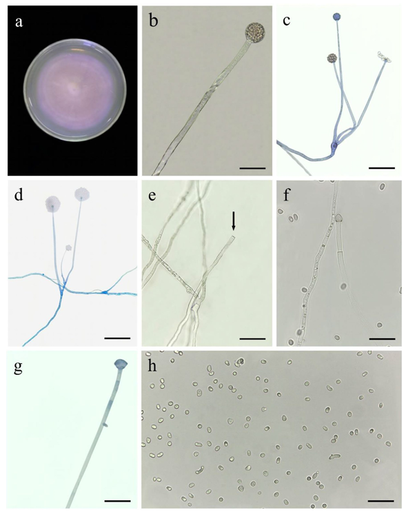
Figure 1 – Microscopic structures of Umbelopsis heterosporus (URM 7882, holotype). a Colony surface after seven days at 25 ºC on PDA. b Simple sporangiophore with globose sporangium. c, d Branched sporangiophore with sporangia. e Simple sporangiophore with inconspicuous columella and collarette (arrow). f–g Simple sporangiophore with columella. h Sporangiospores. Scale bars: 25 μm
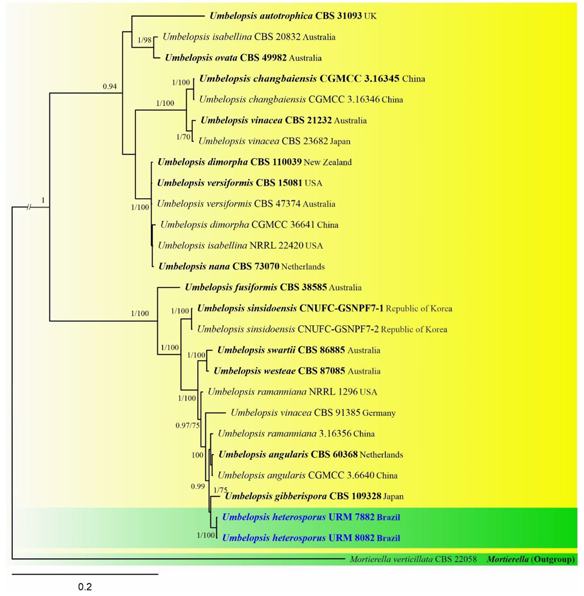
Figure 2 – Phylogram generated from Bayesian inference analysis based on combined LSU and ITS sequence data from 16 representative members of Umbelopsis. Mortierella verticillata (CBS 22058) was used as outgroup. Branches with later Bayesian probabilities (PP) ≥ 0.95 and bootstrap support values for maximum likelihood (ML) analysis ≥ 70 where placed above or below nodes. Ex-type strains are in bold. The newly generated sequences are indicated in blue