Tubulicolla cylindrospora (Morgan-Jones & E.G. Ingram) Réblová & Hern.-Restr., in Réblová, Nekvindová, Kolařík & Hernández-Restrepo, Mycologia 113(2): 420 (2021)
Index Fungorum number: IF 835576; MycoBank number: MB 835576; Facesoffungi number: FoF 15254;
Basionym: Codinaea cylindrospora Morgan-Jones & E.G. Ingram, Mycotaxon 4:504. 1976.
≡ Dictyochaeta cylindrospora (Morgan-Jones & E.G. Ingram) Aramb. & Cabello, Mycotaxon 34:681. 1989. (Nom. inval., Art. 41.4.)
≡ Dictyochaeta cylindrospora (Morgan-Jones & E.G. Ingram) Whitton, McKenzie & K.D. Hyde, Fungal Divers 4:137. 2000.
Description on CMA with Urtica dioica stems – Colonies effuse. Setae absent. Conidiophores 22–68 × (1.5–)2–3.5 μm, up to 6-septate, macronematous, mononematous, sometimes reduced to single conidiogenous cells, unbranched, upright, straight or slightly bent, cylindrical, pale brown toward the base, subhyaline toward the apex. Conidiogenous cells phialidic, 12.5–20.5 × 2.5–4 μm, tapering to (1.5–)2–3.5 μm just below the collarette, narrowly clavate to lageniform to subcylindrical, pale brown to subhyaline becoming hyaline toward the apex, terminal, integrated, with a single apical opening often followed by percurrent elongation of the conidiogenous cell, or with up to four lateral openings formed by successive sympodial elongation. Collarettes 1.5–2 μm diam and 3.5–5 μm deep, funnel-shaped, hyaline, with a narrow, tubular neck. Conidia 8–10.5 × 2–2.5 μm (mean ± SD = 9.3 ± 0.8 × 2.3 ± 0.2 μm), subcylindrical, rounded at both ends, nonseptate, nonsetulate, hyaline, smooth.
Culture characteristics – On CMD: colonies 24–26 mm diam after 28 d, circular, flat, margin entire, velvety, faintly floccose centrally becoming mucoid toward the margin, finely furrowed, colony center whitish becoming beige to brown, creamy toward the margin, reverse beige, brown at the center. On MLA: colonies 30–31 mm diam after 28 d, circular, flat, margin entire to filiform, lanose, funiculose at the inoculation block becoming mucoid, locally cobwebby toward the margin, zonate in the translucent light, colony center whitish becoming dark brown toward the margin, with a prominent beige outer zone of submerged growth, reverse beige-brown. On OA: colonies 30–34 mm diam after 28 d, circular, slightly raised centrally, margin entire, sparsely lanose, funiculose, faintly furrowed becoming cobwebby to mucoid toward the margin, zonate, colony center white-beige becoming brown with several dark brown rings, reverse beige, zonate. On PCA: colonies 15–16 mm diam after 28 d, circular, flat, margin entire, locally cobwebby becoming mucoid-waxy, colony center beige-brown becoming creamy toward the margin, reverse creamy. Sporulation was absent on CMD and MLA, sparse on OA, PCA, and CMA with Urtica dioica stem.
Specimen examined – Cuba, on a fallen leaf of Buchehavia capitata, R.F. Castañeda-Ruiz (INIFAT C94/ 168-1). Culture MUCL 39171.
GenBank Accession Numbers – LSU: EF063575; ITS: MT454494; SSU: MT454478; RPB2: MT454669.
Habitat and distribution – Saprobe on fallen leaves of Buchehavia capitata and Quercus nigra. The species is known from North America in the USA (Alabama) and the Caribbean in Cuba (Morgan-Jones and Ingram 1976; this study).
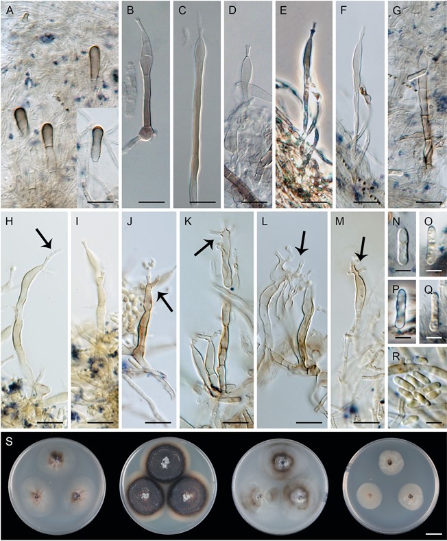
Notes – In vitro, the conidiogenous cells of T. cylindrospora elongate percurrently and sympodially. Percurrent elongation was observed on CMA with Urtica dioica stems, OA (FIG. 15E, H, L), and PCA (Réblová and Seifert 2007:fig. 4c, b). The collarettes form a cascade of openings as a result of up to five percurrent elongations. In older cultures (>8 wk), collarettes sometimes branched dichotomously and conidiogenous cells elongated sympodially to form several lateral openings accumulated at the apex (FIG. 15L). In the CMA culture, we observed clavate, pigmented cells arising from hyaline vegetative hyphae, from which the conidiophores grow (FIG. 15A). The tubular neck between the funnel-shaped collarette and the body of the conidiogenous cell is the prominent character of Tubulicolla. Three morphologically similar Dictyochaeta species, not accepted in this study, can be compared with T. cylindrospora. Dictyochaeta microcylindrospora differs in having smaller (4.8–7.2 × 1–1.5 μm), subcylindrical conidia (Whitton et al. 2000), whereas D. stipitocolla is distinguished in spear-shaped, longer and wider (12.7–17.5 × 2.6–4 μm) conidia with basal protuberances and sterile setae terminated in pointed apical cells (Kuthubutheen and Nawawi 1991b). Dictyochaeta uncinata also possesses a narrow, tubular neck above the body of the conidiogenous cell, but the collarette is not defined, the conidia are longer and narrower (20–25 × 1 μm), uncinate, filiform, and curved, and the setae are sterile with acute tips (Castañeda-Ruiz et al. 1998).

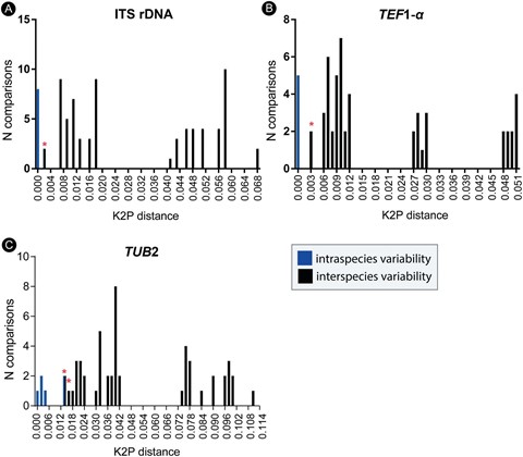
Figure 4. The frequency distribution graphs (bin size of 0.001) of the Kimura 2-parameter distances (barcoding gaps). A. ITS. B. TEF1-α. C. TUB2. The intraspecific distances are shown as blue bars and interspecific distances as black bars. An asterisk (*) marks interspecific variation between D. fuegiana and D. stratosa.

Figure 15. Tubulicolla cylindrospora (MUCL 39171). A. Mycelium and vegetative hyphae with pigmented, thick-walled cells. B–M. Conidiophores (arrows indicate percurrent and sympodial elongations). N–R. Conidia. S. Colonies on CMD, MLA, OA, and PCA after 28 d (from left to right). A–G, N–Q. In CMA with Urtica dioica culture. H–M. In OA culture. Bars: A–M = 10 μm; N–R = 5 μm; S = 1 cm.
