Tomentella dimidiata H.S. Yuan, X. Lu & Y.C. Dai, sp. nov.
Index Fungorum number: IF 555738; MycoBank number: MB 555738; Facesoffungi number: FoF 05614; Figs. 1, 2, 3
Etymology: Refers to the dimitic hyphal structure of the rhizomorphs.
Holotype: IFP 019265.
Basidiocarps annual, resupinate, adherent to the substrate, mucedinoid, without odour or taste when fresh, 0.2–0.4 mm thick, continuous. Hymenophoral surface smooth, golden brown to brown (5D7–6F5) and turning lighter than the subiculum when dry. Sterile margin often determinate, byssoid, concolorous with hymenophore. Rhizomorphs present in subiculum and margins, 12–50 μm diam; rhizomorphic surface more or less smooth; hyphae in rhizomorph dimitic, highly differentiated, of type G (this a new type, see in the notes); central hyphae usually simple septate, thick-walled, unbranched, 2–4 μm diam, pale brown in KOH, cyanophilous, inamyloid; skeletal hyphae from outer part of rhizomorphs thick-walled, unbranched, 1.5–2.5 μm diam, pale brown in KOH, cyanophilous, inamyloid. Subicular hyphae monomitic; generative hyphae clamped, thick-walled, 4–6 μm diam, without encrustation, pale brown in KOH, acyanophilous, inamyloid. Subhymenial hyphae clamped, thin-walled, 4–6 μm diam; hyphal cells short and not inflated, pale brown in KOH, cyanophilous, inamyloid. Cystidia absent. Basidia 10–40 μm long and 4.5–12.5 μm diam at apex, 3.5–5.5 μm at base, with a clamp connection at base, utriform, not stalked, sinuous, without transverse septa, pale brown in KOH and in distilled water, 4-sterigmate; sterigmata 5.5–11.5 μm long and 2–3 μm diam at base. Basidiospores slightly thick-walled, (6.5– )7–7.5(–8)×(5–)5.5–7(–7.5) μm, L=7.07 μm, W=5.99 μm, Q=1.15–1.26 (n=60/2), subglobose to bi-, tri- or quadralobed in frontal and lateral views, echinulate, pale brown in KOH and distilled water, cyanophilous, inamyloid; echinuli usually grouped in 2 or more, up to 2 μm long.
Material examined: CHINA, Liaoning Province, Qingyuan County, Experimental Station of Forest Ecology, on fallen angiosperm branch, 3 August 2016, Yuan 11205 (IFP 019265, holotype); Jilin Province, Changbaishan Nature Reserve, on fallen angiosperm branch, 7 August 2016, Yuan 11267 (IFP 019266).
GenBank numbers: ITS: MK211704, MK211705; LSU: MK446355, MK446356.
Notes: Some species of Tomentella have been reported to have dimitic rhizomorphs, for instance, T. botryoides, T. brunneorufa M.J. Larsen, T. fungicola (Litsch.) M.J. Larsen, T. pilosa, T. punicea (Alb. & Schwein.) J. Schröt. and T. umbrinospora M.J. Larsen (Kõljalg 1996; Daemmrich 2006; Peintner and Dämmrich 2012). In addition, some species of Pseudotomentella Svrček and Odontia Pers. also have dimitic rhizomorphs (Agerer et al. 2001; Tedersoo et al. 2014; Yuan et al. 2018). Agerer recognized six different types (type A–F) of the rhizomorphic hyphal organization, but all these types are monomitic hyphal systems (Agerer 1987–2008). Yorou mentioned the presence of dimitic rhizomorphs in T. punicea and T. umbrinospora, but he treated this kind of structure as type C. However, the dimitic hypal system of rhizomorphs is signifcantly different from the monomitic hyphal structure. We name this kind of structure in rhizomorphs as type G.
Tomentella globospora and T. dimidiata formed a clade in the phylogenetic tree (Fig. 4), and they share the similar morphological and anatomical characteristics: basidiocarps adherent to the substrate, thick-walled subicular hyphae, utriform basidia and the absence of cystidia. However, T. globospora differs from T. dimidiata by having crustose basidiocarps, globose to subglobose basidiospores and the absence of rhizomorphs. The key feature of T. dimidiata is the presence of dimitic rhizomorphs in subiculum. T. incrustata and T. dimidiata both have a byssoid sterile margin, short subhymenial hyphal cells, and subglobose to bi-, trior quadra-lobed basidiospores; but T. incrustata has clavate basidia, encrusted subicular hyphae and generative hyphae occasionally inflated.
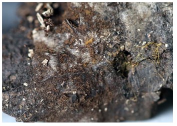
Figure 1 –A basidiocarp of Tomentella dimidiata (IFP 019265, holotype)
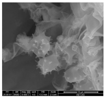
Figure 2 – SEM of basidiospores of Tomentella dimidiata (IFP 019265, holotype)
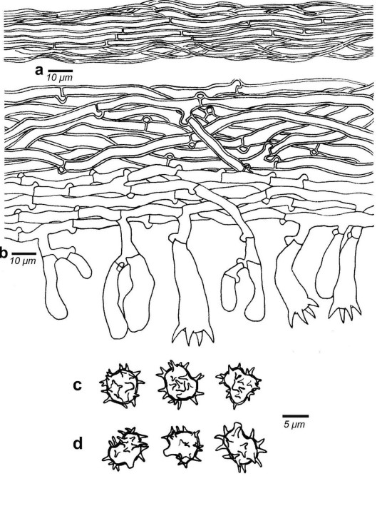
Figure 3 – Microscopic structures of Tomentella dimidiata (IFP 019265, holotype). a Hyphae from a rhizomorph. b Section through a basidiocarp. c Basidiospores in frontal view. d Basidiospores in lateral view
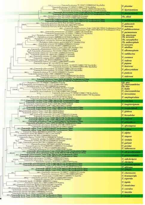
Figure 4 – Phylogenetic position of Tomentella species inferred from the ITS sequences. Bootstrap support values for ML≥50% and Bayesian posterior probabilities≥0.95 are given near nodes respectively. The tree is rooted with Odontia fibrosa (TU115714). The new species are in bold