Sulcosporium thailandica Phookamsak & K.D. Hyde.
Index Fungorum number: IF623003; Facesoffungi number: FoF00895; Fig. 1
Etymology – The generic epithet “thailandica” refers to the country where the fungus was first collected.
Holotype – MFLU 11-0243
Pathogen on Axonopus compresus (Sw.) P. Beauv., causing necrotic leaf spots. Lesions 3 – 5 cm long, usually forming from leaf margins, irregular in shape, visible as pale brown to brown regions, separated from healthy part of leaf by reddish brown margins. Sexual morph: Ascomata 70 – 130 μm high, 80 – 140 μm diam., solitary, scattered, immersed, globose to subglobose, uni-loculate, membranous, with minute central papilla erumpent through host surface, ostiolate. Peridium 8 – 20 μm wide, composed of several layers of thick-walled, brown to dark brown, pseudoparenchymatous cells, arranged in a textura angularis. Hamathecium comprising dense, 2 – 4 μm wide, cellular pseudoparaphyses, constricted at the septum, with distinct septa, anastomosing at the apex, embedded in mucilagenous matrix . Asci ( 7 0 – ) 80 – 100 (−110) × (24.5–) 27 – 33 (−36) μm (x̄ = 86.2 × 30.4 μm, n = 25), 8 – spored, bitunicate, fissitunicate, broadly fusiform to clavate, saccate or ampulliform, with a short blunt pedicel, apically rounded, with well-developed ocular chamber. Ascospores (27–) 29 – 35 (−37) × 8 – 10 μm (x̄ = 33.4 × 9.8 μm, n=30), overlapping bi- to tri-seriate, initially hyaline, becoming very pale brown, ellipsoidal to fusiform, or slightly clavate, with rounded ends, 1-septate, not constricted at the septum, rough, furrowed, thick-walled, slightly swollen above septa. Asexual morph: Undetermined.
Culture characteristics – Colonies on PDA slow growing, 20 – 28 mm diam. after 4 weeks at 25 – 30 °C, colonies irregular, sparse to mediun dense, flattened, slightly raised, smooth with fimbriate to rhizoid edge, thinly hairy to woolly, smooth, from above dark greenish to dark brown at the margin, grey to dark grey in the centre; reverse dark brown at the margin, black at the centre, not producing pigment in PDA.
Material examined – THAILAND, Chiang Rai Province, Muang District, Khun Korn Waterfall, on living leaves of Axonopus compresus (Poaceae), 21 June 2011, R. Phookamsak RP0125 (MFLU 11-0243, holotype), ex-type living culture, MFLUCC 12-0004, BCC.
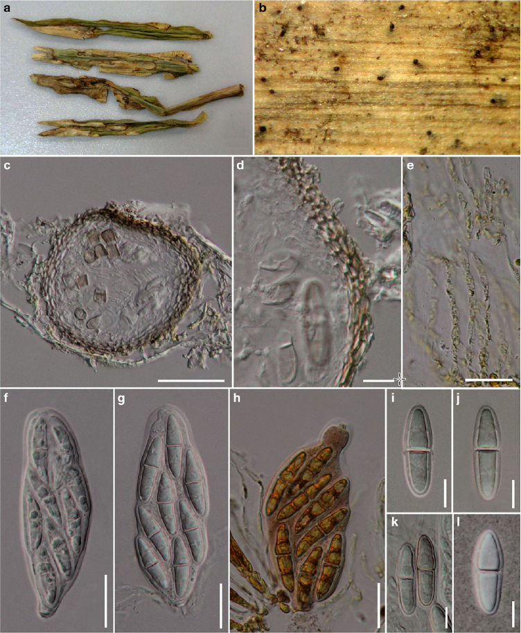
Fig. 1 Sulcosporium thailandica (holotype) a Lesions on host leaves b Papilla of ascomata visible as black spots on host surface c Vertical section through an ascoma d Section through peridium e Pseudoparaphyses stained in Melzer’s reagent f, g Asci h Ascus stained in Melzer’s reagent i – k Ascospores l Ascospore stained in Indian ink. Scale bars: c = 50 μm, e – h = 20 μm, d, i, j, k, l = 120 μm.