Subulicystidium daii S.L. Liu & L.W. Zhou
MycoBank number: MB 559882; Index Fungorum number: IF 559882; Facesoffungi number: FoF 12862;
Description
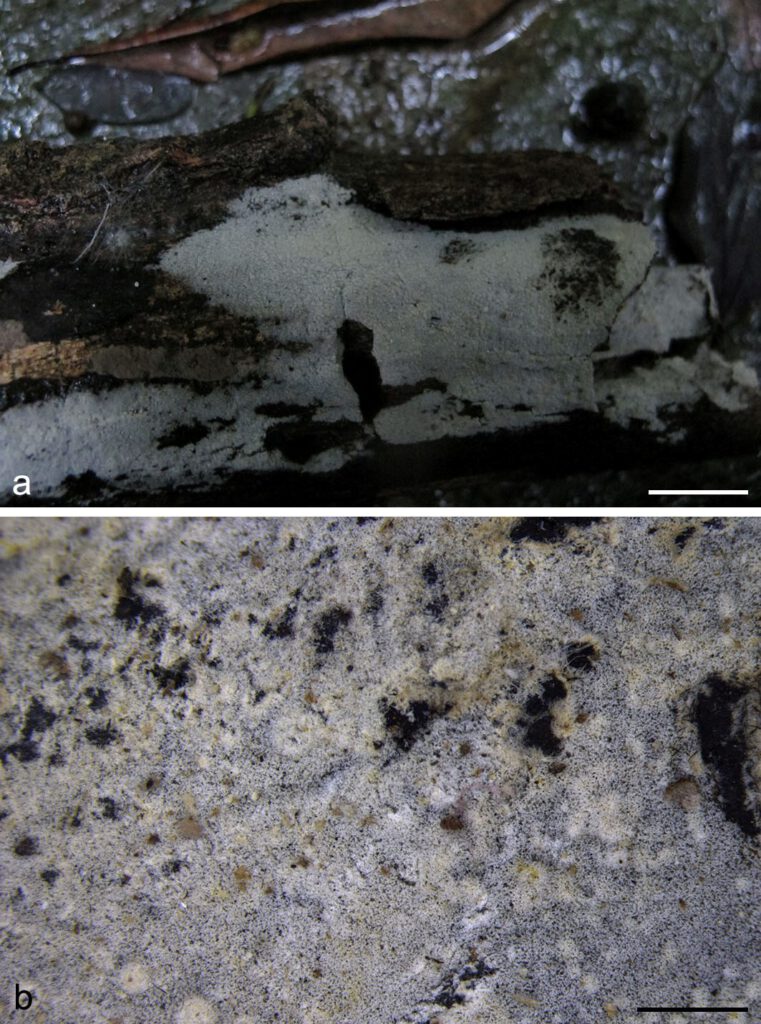
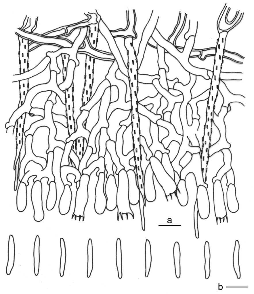
Basidiomes annual, resupinate, effused, very thin, loosely attached to the substrates, up to 9 cm long, 4 cm wide. Hymenophore smooth, cream to straw-yellow when fresh, cream to ash-grey with age, not cracked. Margin undifferentiated.Hyphal system monomitic; generative hyphae with clamp connections, hyaline, slightly thick-walled, frequently branched and septate, loosely subparallel, 2–3.5 µm in diam. Cystidia abundant, subulate, projecting beyond hymenium, hyaline, thick-walled, regularly covered with rectangular crystals except at the apex, 50–80 × 3–5 µm. Basidia subclavate to suburniform, hyaline, thin-walled, with four sterigmata and a basal clamp connection, 16–22 × 5–7 µm; basidioles in shape similar to basidia, but slightly smaller. Basidiospores fusiform to slightly vermicular, hyaline, thin-walled, smooth, inamyloid, indextrinoid, acyanophilous, (15–)15.5–17.5(–18.5) × 2.3–3 µm, L = 16.5 µm, W = 2.6 µm, Q = 6.5–6.9 (n = 60/2).
Material examined: CHINA, Hubei, Wudangshan Town, Wudangshan National Forest Park, on fallen angiosperm branch, 20 Aug. 2017, L.W. Zhou, LWZ 20170820-35 (holotype in HMAS).
Distribution: CHINA
Notes: Besides Subulicystidium acerosum, S. daii also resembles S. cochleum and S. perlongisporum by the long (above 15 µm in length) and straight or slightly curved basidiospores; however, S. cochleum differs in the presence of a bundle of needle-like crystals at the cystidial crystalline sheath ends, while S. perlongisporum differs in narrower basidiospores (1.5–2.5 µm in width; Ordynets et al. 2018).

Fig. 1. Basidiomes of Subulicystidium daii (LWZ 20170820-35, holotype). — Scale bars: a = 1 cm; b = 1 mm.

Fig. 2. Microscopic structures of Subulicystidium daii (drawn from the holotype). a. Vertical section of basidiomes; b. Basidiospores. — Scale bar = 10 μm.
