Sublophiostoma thailandica Phookamsak, Hongsanan, Goonas. & K.D. Hyde, sp. nov.
MycoBank number: MB 557973; Index Fungorum number: IF 557973; Facesoffungi number: FoF 09403 (Figs. 3, 4).
Etymology: The specific epithet “thailandica” refers to the country from which the species was collected.
Holotype: MFLU 11–0158.
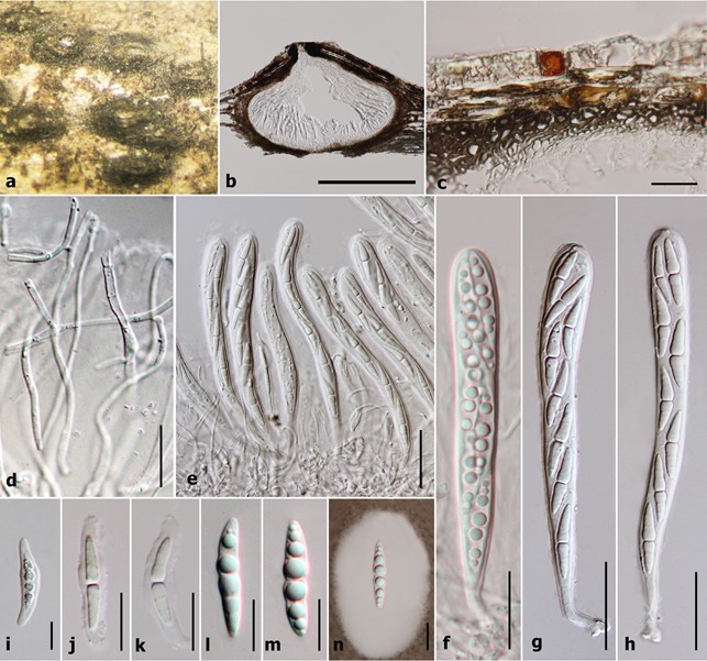
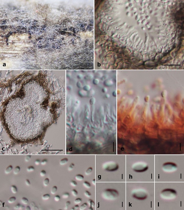
Saprobic on grasses and bamboo (Poaceae). Sexual morph: Ascomata 450–600 µm high × 135–185 µm diam. (x̅ = 505 × 160 µm, n = 10), solitary to gregarious, scattered, immersed to semi-immersed or erumpent, visible as dark ellipsoidal spots on host surface, uni-loculate, coriaceous, black, subglobose, hemisphaerical to lenticular, glabrous, central ostiole with minute papilla beneath the host tissue, crest-like opening. Peridium 13–30 µm wide, thick at upper lateral part and thinner at the base, composed of several layers of cells, with outer layers of thick, dark brown layers of cells of textura angularis and inner layers comprising hyaline, somewhat flattened cells of textura angularis to textura epidemoidea. Hamathecium comprising 0.6–1.5 µm wide, dense, septate, branched trabeculate pseudoparaphyses, embedded in a gelatinous matrix. Asci 85–105 µm × 8.5–10.5 µm (x̅ = 95 × 9 µm, n = 30), 8-spored, bitunicate, fissitunicate, cylindric-clavate, rounded at apex, with a small bulbous pedicel, with well-developed ocular chamber. Ascospores 20.4–25.5 × 3.5–5.6 µm (x̅ = 23.7 × 4.5 µm, n = 30), uni to bi-seriate, partially overlapping, hyaline, fusiform, 1-septate, constricted at the septum, each cell wider near the septum, becoming acute at the ends, guttulate, smooth-walled, surrounded by a large mucilaginous sheath. Asexual morph: Coelomycetous, produced on bamboo pieces on WA after 8 weeks, appearing as black, punctiform, globose structures, covered by grey to dark grey vegetative mycelium. Conidiomata solitary or aggregated, pycnidial, obpyriform. Conidiomata walls 5–10 µm (x̅ = 7 µm, n = 14) in thickness, comprising outer, brown cells of textura globulosa to textura angularis and inner, hyaline cells of textura angularis. Conidiophores reduced to conidiogenous cells. Conidiogenous cells 5.7–7.7 µm (x̅ = 6.7 µm, n = 13) in length, phialidic, with narrow peri- clinal thickening, hyaline, integrated. Conidia 1.8–2.6 × 1.1–1.8 µm (x̅ = 2.4 × 1.4 µm, n = 20), hyaline, aseptate, smooth-walled, subglobose to oblong, with large guttules. Colonies on PDA medium showing slow growth, 30–35 mm diam. after 4 weeks at 25–30 °C, white to pale yellow at the magins, white to grey in the center, with dark grey turfing in the middle of the colony; reverse white at the outer margin, more grey towards the inner area, becoming yellowish-grey in the center, dense, irregular, slightly raised to umbonate, dull with undulate edge, velvety, not produced pigmentation on agar.
Material examined: THAILAND, Chiang Mai Prov., Chom Tong District, Doi Inthanon, on dead stems of grass, 16 November 2010, R. Phookamsak RP0038 (MFLU 11–0158, holotype), ex-type living culture, MFLUCC 11–0207, ibid. on dead branches of bamboo, 6 October 2010, R. Phookamsak, RP0081 (MFLU 11–0201), living culture MFLUCC 11–0165; on dead stem of Thysanolaena maxima Kuntze, 6 October 2010, R. Phookamsak, RP0088 (MFLU 11–0208), living culture, MFLUCC 11–0172; on dead stem of Thysanolaena maxima, 16 November 2010, R. Phookamsak. RP0090 (MFLU 11–0210), living culture MFLUCC 11–0174; on dead stem of Thysanolaena maxima, 16 October 2010, R. Phookamsak, RP0101 (MFLU 11–0221), living culture, MFLUCC 11–0185; Chiang Rai Prov., Muang District, Khun Korn Waterfall, on dead stem of Thysanolaena maxima, 21 June 2011, R. Phookamsak, RP0128 (MFLU 11–0245), living culture MFLUCC 12–0006.

Figure 3. Sexual morph characters of Sublophiostoma thailandica MFLU 11–0158. (a) Appearance of ascomata on host. (b) Section of ascoma. (c) Peridium. (d) Trabeculate pseudoparaphyses. (e–h) Asci. (i–m) Ascospores. (n) Appearance of mucilaginous sheath surrounding young ascospore (stained in Indian ink). Scale Bars: b = 100 µm, c, d = 20 µm, e–i, l–n = 10 µm, j, k = 5 µm.

Figure 4. Asexual morph characters of Sublophiostoma thailandica MFLU 11–0158. (a) Conidiomata on bamboo pieces on WA after 8 weeks. (b) Conidioma wall. (c) Section of conidioma. (d) Conidiogenous cells. (e) Conidiogenous cells stained in congo red. (f–l) Conidia. Scale bars: b = 10 µm, c = 50 µm, d, e, f = 2 µm, g–l = 1 µm.
