Stromatoneurospora phoenix (Kunze ex Fr.) S.C. Jong and E.E. Davis, Mycologia 65: 459 (1973),Figure 1.
Index Fungorum number: IF 324283; MycoBank number: MB 324283; Facesoffungi number: FoF 03077;
Sexual morph: Stromata scattered on the host surface, subglobose to ovate, stipitate, roughened, 2–6 mm diameter, stipes short, slender, unbranched, smooth, black, 2–2.2 mm long, deeply rooting in the substrate; externally Tawny Blended (8), Umber (9) or Apricot (42) with black papillate ostioles of embedded perithecia, internally white. Texture fairly hard, lacking carbonaceous layer. Perithecia completely immersed beneath the stroma, surface obovoid to globose, 650–850 × 750–100 µm; ostioles conspicuous, black, papillate. Paraphyses typical of the stromatic Xylariales, tapering, 3–5 septate, (200–) 300–325 × 5–7.5 µm (x̄ = 283.69 × 5.97 µm; n = 50). Asci unitunicate, eight-spored, cylindrical, (150) 175–200 × (7.5–) 10–12.5 µm. Ascospores ellipsoid-fusiform, 1-celled, hyaline at first, becoming yellow-brown and black in maturity, (15–)18–20(–21) × (7–)8–9(–10) µm, (x̄ = 18.7 × 8 µm; n = 50) with longitudinal, parallel to convergent, continuous to discontinuous ridges on the wall resembling those of Neurospora ascospores and have neither germ pore nor germ slits. Asexual morph: Lindquistia-like. Synnemata cylindrical to clavate, 24–25 × 2–3 mm. Conidiophores loosely arranged, branched, undetermined in length, 2–3 µm diameter. Conidiogenous cells produced holoblastically, cymbiform, rarely subglobose to obovoid, hyaline, 9–10 × 4–5 µm, each cell producing one or several conidia. Conidia hyaline, smooth, subglobose, obovoid, ellipsoid with flattened base, 4–6 × 2–3 µm.
Culture characteristics – Colonies on OA reaching the edge of a 9 cm Petri dish in 1 week, at first whitish becoming velvety to felty, azonate with entire margin, peach (4), flesh (37), or salmon (41) after 1 month incubation (Figure 1g,h). Synnemata produced after 1 month of incubation at room temperature (ca. 20–25 ◦C; Figure 2a,b). Colonies on YMGA covering Petri dish in a week, at first whitish, becoming peach (4), flesh (37), and salmon (41), velvety to felty, azonate with entire margin.
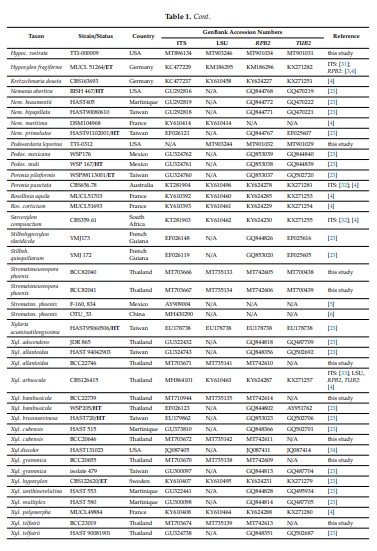
Materials studied – Thailand, Chiang Mai Province, Ban Hua Thung community forest, 19.42044j N, 98.97140j E, on burnt grass, 6 July 2016, P. Srikitikulchai, S. Wongkanoun, BBH 42282, corresponding cultures BCC82040 and BCC82041, independently obtained from two different stromata of BBH-42282); GenBank accession numbers for DNA sequences are presented in Table 1.
Notes –The morphological characteristics of our fungus are clearly similar to those of the holotype of Stromatoneurospora phoenix that was reported from Surinam, as well as to specimens that were later reported from Brazil, Puerto Rico, and the USA. Aside from S. phoenix there is only one other species that was assigned to the genus, i.e., Stromatoneurospora elegantissima, reported from burnt grass in Brazil. In keeping with the description of Jong and Davis [2], this species differs from S. phoenix in having much larger ascospores (25 × 12 µm). Three other genera are morphologically similar to Stromatoneurospora by having a fairly hard stromatal texture, lacking a carbonaceous layer, and some of them also produce a lindquistia-like anamorph: Podosordaria, Poronia, and Sarcoxylon also show affinities with Stromatoneurospora but differ in their ascospore morphology.


Figure 1. Morphological characteristics of Stromatoneurospora phoenix (specimen BBH 42282). (a,b): stromata in the natural habitat; (c): stromatal surface and ostioles; (d): longitudinal section of stroma showing perithecia and the tissue below the perithecial layer; (e): asci with apical apparatus bluing in Melzer’s reagent (black arrow); (f,g): ascospores by scanning electron microscopy (SEM); (h–k): ascospores by light microscopy. Scale is indicated by bars ((a): 2 mm. (b): 1 mm. (d): 500 µm; (e): 20 µm, (f–k): 5 µm).
