Srinivasanomyces kangrensis S. Rana & S.K. Singh, sp. nov.
MycoBank number: MB 830718; Index Fungorum number: IF 830718; Facesoffungi number: FoF 06589; Fig. 100
Etymology: The specific epithet refers to the place of collection
Holotype: AMH-10175
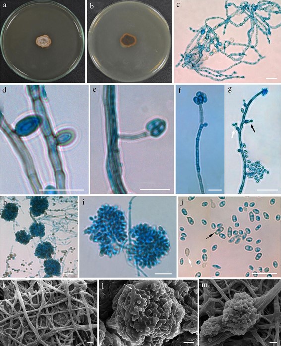
Isolated from dead bark of Prunus cerasoides. Sexual morph Undetermined. Asexual morph Hyphae thin, simple to branched, sometimes forming bundles, smooth-walled, subhyaline, septate, sometimes constricted near septa, 0.9–3.1 µm ( x̄ = 1.8 µm, n = 30) wide. Chlamydospores abundant, solitary or in chains, globose to subglobose, smooth-walled, pigmented, subhyaline to light olivaceous, 3.1–7.8 × 2.4–5.7 µm ( x̄ = 5.2 × 3.9 µm, n = 30). Produces multiple morphs (synanamorphs) of Diplococcium and Phialocephala. Conidiophores produced from lateral hyphae, indeterminate, intercalary, simple to densely branched, septate, smooth-walled, subhyaline to light olivaceous. Conidiogenous cells subhyaline to light olivaceous, lateral to terminal, phialidic, densely produced, monoblastic to polyblastic. Phialides variable, solitary or in dense clusters forming globose conidial heads 12.3–45.9 × 8.1–37.6 µm ( x̄ = 18.7 × 16.1 µm, n = 30), subhyaline to light olivaceous, smooth-walled, collarette, cylindrical to ampulliform, 3.7–58.5 × 0.8–3 µm ( x̄ = 13.7 × 1.4 µm, n = 30). Conidia produced in gleosporic mass, sometimes directly from superficial lateral hyphae, width 1.9–2.3 µm (x̄ = 2.1, n = 10), subhyaline to light olivaceous, smooth-walled, pyriform to obpyriform, globose to subglobose, fusoid, clavate, solitary to catenate, 1–2 guttules.
Culture characteristics: Colonies on PDA reaching to 15–20 mm diam. after 3 weeks at 25 °C; colonies from above greyish white (7B1) to fawn/light brown (7D4), flat, circular, margin irregular smooth, slightly cottony; colony from reverse, dark brown (8F4).
Material examined: INDIA, Himachal Pradesh, Kangra District, Simbal (31.9754 N” 76.6507 E”), from dead bark of Prunus cerasoides, 10 April 2018, S. Rana, AMH 10175 (holotype), ex-type living culture, NFCCI 4505.
GenBank numbers: ITS =MK478471, LSU = MK478470.
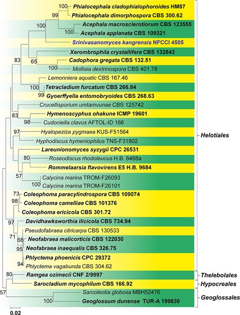
Notes: Srinivasanomyces kangrensis differs from other taxa based on the sequence analysis as well as morphology (Figs. 100, 101). In the megablast analysis, an ITS sequence of S. kangrensis, was 88.03% (368/418) similar with 6 gaps (1.4%) with Phialocephala cladophialophoroides Madrid et al. (HM87, type), 87.63% (418/477) similar with 7 gaps (1.4%) with P. dimorphospora W.B. Kendr. (CBS 300.62, type), 87.86% (420/478) similar with 7 gaps (1.4%) with Acephala applanata Grünig & T.N. Sieber (109321, type) and 86.84% (416/479) similar with 9 gaps (1.87%) with A. macrosclerotiorum Münzenb. & Bubner (CBS 123555, type). Srinivasanomyces kangrensis differs from Phialocephala oblonga (C.J.K. Wang & B. Sutton) Tanney, Seifert & B. Douglas (Tanney et al. 2016) in having dense globose clusters of conidial heads, while in P. oblonga, conidia are produced laterally from compact cylindrical synnemata. Phialocephala dimorphospora (Kendrick 1961) produces conidiophores from lateral superficial hyphae forming penicillate heads, which bears numerous phialides, while the conidiophores of Srinivasanomyces kangrensis are indeter- minate, intercalary, in simple to dense globose to subglobose clusters. In Phialocephala catenospora Tanney & B. Douglas (Tanney et al. 2016) dark brown septate conidia are produced from short lateral stalks in long branched catenate chains and from solitary or groups of phialides. Phialocephala nodosa Tanney & B. Douglas (Tanney et al. 2016) produces irregular dark brown microsclerotia. The phialides of P. aylmerensis Tanney & B. Douglas (Tanney et al. 2016) are produced as solitary loose clusters, while conidiophores of P. victorin Vujan. & St-Arn. (Vujanović et al. 2000) arise singly or in groups, are mono- to tripenicillate, and form terminally or laterally on the hyphae. Proliferated phialides have an extremely expanded collarette.

Fig. 100 Srinivasanomyces kangrensis (AMH 10175, holotype). a Colony morphology on PDA (front view). b Reverse view of colony. c Vegetative hyphae and chlamydospores. d Conidia produced from lateral hyphae. e, f Conidiophores produced from lateral hyphae with conidia. g Arrow showing conidia produced from phialide. h Tufts of slimy gleosporic mass of conidia with densely produced phial- ides. i Magnified view of gleosporic mass of conidia (arrow showing reduced phialides). j Conidia (arrow shows truncate and oblique base). k SEM of hyphae. l SEM of magnified view gleosporic mass of conidia (arrow showing densely produced phialides). m SEM of tufts of slimy gleosporic mass of conidia. Scale bars: c–j = 20 μm, k–m = 5 μm

Fig. 101 Phylogram generated from Maximum likelihood analysis for Srinivasanomyces kangrensis NFCCI 4505 (AMH 10175) using combined ITS and LSU sequence data based on the Tamura-Nei model (Tamura and Nei 1993). Bootstrap support values are indicated at the nodes and values below 50% are not shown. Phylogenetics analyses were conducted in MEGA7 (Kumar et al. 2016). The novel taxon is shown in blue colour and type taxa used are depicted in bold
