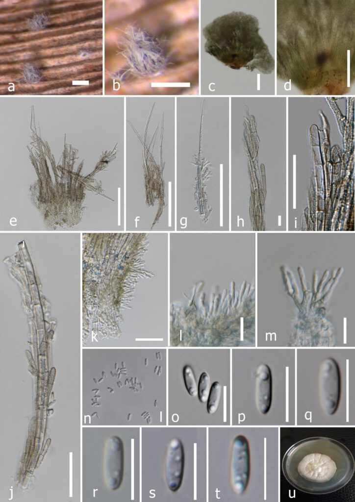
Smaragdiniseta musae Samarakoon & Chomnunti, sp. nov. Figure 02
MycoBank number: MB 844095; Index Fungorum number: IF 844095; Facesoffungi number: FoF 10846;
Etymology: The species epithet reflects the host genus, Musa
Holotype: MFLU XXXX
Saprobic on dead leaves of Musa sp. Sexual morph: Undetermined. Asexual morph: Sporodochia 0.2–0.35 mm diam., cup-shaped, scattered, solitary, circular with an entire margin, initially emerald green, later becoming black, composed of hyaline, erect conidiophores, surrounded by filamentous white marginal hyphae and setae at periphery. Fungal hyphae parallelly arranged like a palisade layer, compacted, verrucose, olivaceous brown or hyaline, sometimes warticulate, septate, branched, irregularly thick-walled, with ultimate hyphal cell rounded at apex and flat at base; hyphal cells: 10–14×0.8–2.5μm (x̄=13.3×1.6μm, n=30). Aggregated hyphae hyaline or emerald green when immature, tan brown at maturity. Setae fast growing from marginal hyphae, numerous, aggregating as a pale gray brush, elongated, straight or slightly curved, tapering towards apex, hyaline, septate, unbranched, apiculate at apex, sometimes caudate, truncate or rounded, swollen to globose to ovate or rounded at base, setae wall 0.4–1.5μm (x̄=0.8μm, n=30) thick, often smooth, sometimes rough with hyaline acellular coatings, 60–250×0.8–2.8μm (x̄=126.4×1.6μm, n=30); septa 4–8μm apart. Marginal hyphae often coiling or growing around the setae or projecting out, forming a wefty cover around setae, 95–125μm (x̄=106μm, n=30) wide. Conidiophores 4–14×1.3–2.5μm (x̄=12.9 ×1.7μm, n=30), macronematous, hyaline, smooth, thin-walled, arising from sub hyaline or pale brown, with slightly thickened and swollen basal cells, often with a narrow truncate base, wider in the middle, tapering to rounded at apex, 3–4×5–6μm (x̄=3.5 ×5.5μm, n=10), septate, rarely unbranched, mostly branched and with 2–3 conidiogenous sub-branches at each node. Conidiogenous cells phialidic, rough or thin-walled, rod-shaped, elongated or ovate, 4–9×1–3μm (x̄=6.8 ×1.9μm, n=20), without a collarette, pinching off simple conidia at the apex of each phialide. Conidia simple, hyaline, smooth, thin-walled, elliptic or slightly ovate, rounded at one end and acute at other end, with two distinct guttules at vertical ends, sometimes with 3–5 guttules, 6–10×2–3.5μm (x̄=8.3×2.7μm, n=10).
Culture characteristics: Conidia germinating on PDA after 48 hours, germ tubes were produced from the acute end. Colonies growing on PDA reaching 20 mm diam. after 2 weeks in the light conditions at 25 °C, mycelium mostly immersed, not slimy, cottony, pinkish-white, dense in the middle and comparatively sparse at the periphery. Radially and unevenly striated, colonies have a slightly wrinkled appearance from the top. The formation of sporodochia was not observed in mature colonies.
Material examined –THAILAND, Chiang Rai Province, Mae Sai, on a dead leaf of Musa sp., 16 June 2019, Binu Samarakoon, BNS264 (MFLUXXXX holotype), living cultures (BNS264A)MFLUCC XXXX, (BNS264B) MFLUCC XXXX.
Notes: Based on BLASTn search results of ITS, tub2, rpb2 sequence data, Smaragdiniseta musae showed a high similarity (ITS=97.23%, tub2=89.16% and rpb2=91.10%) to S. bisetosa (CBS 459.82). In the multigene phylogeny, S. musae clustered with S. bisetosa in having strong statistical support (100% ML, 1.00 BYPP) (FIGURE 1). The base pair comparison of ITS, tub2 and rpb2 of our new taxon revealed 3.14% (17/540), 12.36% (35/283) and 9.34% (63/674) nucleotide differences with S. bisetosa. Besides, S. musae differs from S. bisetosa by the conidial morphology. The conidia of S. musae have two distinct guttules at the vertical poles. In addition, some conidia bear minute guttules at the center. However, the taxonomic illustration and description of S. bisetosa did not indicate the guttule formation in the conidia (Rao & Hoog 1983). Moreover, the conidia of S. bisetosa are obclavate, narrowly ellipsoidal or rod-shape, whereas our new taxon has an elliptic or slightly ovate shaped conidia with a rounded top and an acute base. Both ends of the conidia of S. bisetosa are rounded or sometimes are found with a truncate base (Rao & Hoog 1983). We have not observed a truncate base in the conidia of S. musae. Furthermore, the marginal hyphae of S. musae often coil or grow around the setae but never have over grown. The marginal hyphae always reach around 95–125μm of the setae and terminate at a point. According to the description of Rao & Hoog (1983), in S. bisetosa, the marginal hyphae always covered the entire setae. Our new collection is similar in morphology to the other genera in Stachybotryaceae in having conidiophores where the ultimate branches become phialides (Lombard et al. 2016). This feature phenotypically justifies the placement of our new collection in Stachybotryaceae. Based on distinct morphological characteristics and strong statistical support from our molecular phylogeny, Smaragdiniseta musae is therefore herein introduced as a new species on Musa sp. from Chiang Rai Province, Thailand. This is the first report of Smaragdiniseta on Musaceae and also from Southeast Asia. In addition, S. musae is the second taxon that is being described in this genus.

FIGURE 2. Smaragdiniseta musae (MFLU XXXXX, holotype) a, b Sporodochia on the host c d Cupulate sporodochia e-j Setae and marginal hyphae k-m Attachments of conidiophores and phialides n-t Conidia o Colony on PDA after 8 weeks. Scale bars: a, b=400μm, c, d, i, j=100μm, e-h =50μm, k=20μm, l-t=10μm.
