Similitrichoconis wongii R.F. Castañeda, M. Vera & D. Sosa, sp. nov. Figs 2–5
MycoBank number: MB 833731; Index Fungorum number: IF 833731; Facesoffungi number: FoF;
Differs from Trichoconis spp. by its monoblastic determinate conidiogenous cells and the production of obclavate conidia after schizolytic conidial secession.
Type: Ecuador, Los Ríos province, Cantón Buena Fé, Parroquia Patricia Pilar, 0°35’45″S 79°21’49″W, on submerged decaying leaf of an unidentified plant in stream, 18.III.2019, coll. A. Quevedo, M. Vera & F. Espinoza (Holotype, CCMCIBE-H641).
Etymology: wongii (Latin) The new species is dedicated to Vicente Wong who provided permission and facilities for sample collection in the Rio Palenque reserve belonging to the Wong Foundation
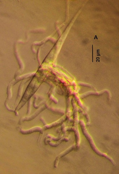
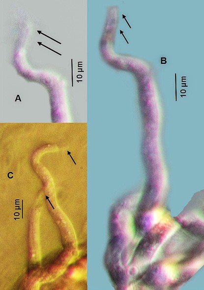
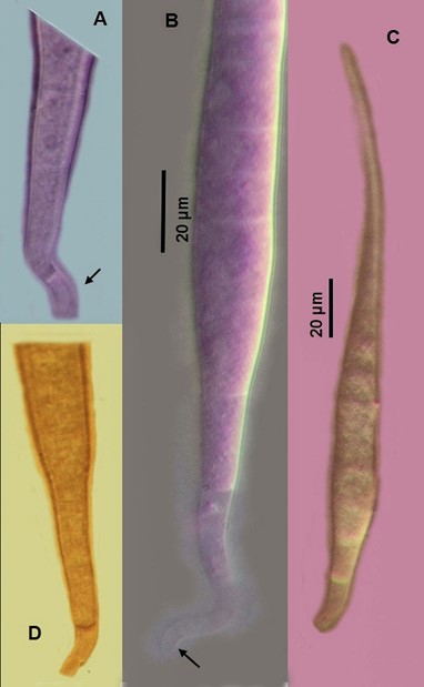
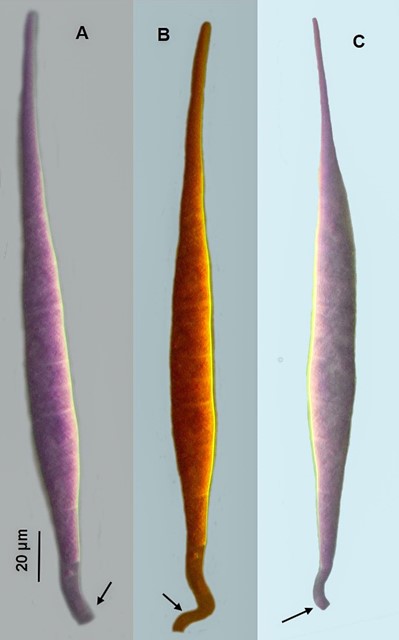
Colonies on the natural substrate effuse, hypophyllous, funiculose, white. Mycelium mostly superficial and immersed; hyphae septate, branched, smooth, 3–5 µm diam, hyaline. Conidiophores macronematous, mononematous, erect or prostrate, flexuous, sinuous, unbranched, 3–5-septate, smooth, hyaline, 45–80 × 6–9 µm, with 2(–3)-cells clear to translucent toward the apex. Conidiogenous cells monoblastic, cylindrical to slightly subulate, strongly curved to uncinate, integrated, determinate, terminal, smooth, clear to translucent, 8–10 × 3–5 µm. Conidial secession schizolytic. Conidia solitary, acrogenous, obclavate, long fusiform, 8–15-euseptate, rostrate toward the apex, rostrum ≤10 µm long, smooth-walled, hyaline, 200–250 × 10.5–14 µm, with a conspicuous, curved to uncinate, clear to translucent basal cell, 2–4 µm long.
Notes: Similitrichoconis wongii presents two clear to translucent cells, the conidiogenous cell and (after schizolytic secession) a conidial basal cell. Both clear to translucent cells could be interpreted either as a 2-celled isthmus or two separating cells; unfortunately despite several attempts we obtained no culture that would clarify this uncommon conidiogenous event. The obclavate conidia with a cylindrical hyaline basal cell in S. wongii somewhat resemble Trichoconis spp. However, the basal frill observed in Trichoconis originates after rhexolytic secession of a narrow cylindrical separating cell produced at several loci of the uniformly colored polyblastic conidiogenous cells (Braun & al. 2016, Silva & al. 2016). The cylindrical denticles and pedunculate cells in Pendulispora venezuelanica M.B. Ellis (illustrated by Ellis 1961) are superficially similar to the conidial basal cells and conidiogenous cells in S. wongii, but the evident sympodial extension of the conidiogenous cells are typical of polyblastic conidiogenous cells (Seifert & al. 2011). Ellis (1971), who ignored these sympodial extensions of the conidiogenous cells, described the conidiogenous cells as monoblastic. The existence of hyaline separating cells during and after conidiogenous events occurs in several hyphomycete genera (e.g., Arcuadendron Sigler & J.W. Carmich., Coccidioides C.W. Stiles, Chrysosporium Corda, Malbranchea Sacc., Ovadendron Sigler & J.W. Carmich.), but there rhexolytic conidial secession occurs during the conidial arthro-thallic disarticulation (Sigler & Carmichael 1976, Seifert & al. 2011).

Fig. 2. Similitrichoconis wongii (ex-holotype, CCMCIBE-H641). Colony from natural substratum.

Fig. 3. Similitrichoconis wongii (ex-holotype, CCMCIBE-H641). A. Conidiogenous cell; B, C. Conidiophores and conidiogenous cells. (Arrows indicate clear (A,C) and translucent (B) conidiogenous cells.)

Fig. 4. Similitrichoconis wongii (ex-holotype, CCMCIBE-H641). A, D. Lower conidial section above stained translucent basal cells. B. Lower conidial section with curved clear basal cell. C. Conidium with translucent basal cell.

Fig. 5. Similitrichoconis wongii (ex-holotype, CCMCIBE-H641). A–C. Conidia with curved translucent basal cells (indicated by arrows) following schizolytic conidial secession.
