Seriascoma bambusae H.B. Jiang, K.D. Hyde & Phookamsak, sp. nov.
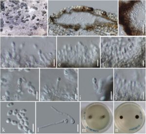
Index Fungorum number: IF558430; Facesoffungi number: FoF 09885; Figure 4
Etymology: The specific epithet “bambusae” refers to the host, bamboo, on which the new species was collected.
Holotype: KUN–HKAS 112014
Saprobic on dead culms of bamboo. Sexual morph: Undetermined. Asexual morph: Coelomycetous. Conidiomata 170–380 μm diam., 110–150 μm high, solitary to gregarious, immersed under the host’s cortex, raised, becoming superficial, dull, black, elongate–conical to lenticular or dome–shaped, uni– to bi–loculate, glabrous. Locules 95–220 μm diam., 35–140 μm high, clustered, dark brown to black, subglobose. Peridium 10–35 μm thick, thin– to thick–walled, of unequal thickness, thick at the sides, thin at the base, composed of host and fungal tissue, with several layers of dark brown to black, pseudoparenchymatous cells of textura angularis. Conidiophores reduced to conidiogenous cells. Conidiogenous cells 5.6–7.2 × 1.6–3.5 μm (x̅ = 6.4 × 2.5 μm, n = 20), enteroblastic, phialidic, determinate, discrete, cylindrical to ampulliform, hyaline, aseptate, smooth–walled. Conidia 3.5–4 × 2–2.3 μm (x̅ = 3.8 × 2.2 μm, n = 20), subglobose to ellipsoidal, hyaline, 2–guttulate, aseptate, smooth–walled.
Culture characteristics: Conidia germinating on PDA within 24 hours. Colonies were growing slowly on PDA, reaching 5 mm in one week at room temperature (10–20 °C), under normal light conditions, colonies cottony, circular, raised, greyish to dark brown from above and below. Mycelium superficial or immersed in media, with branched, septate, smooth hyphae.
Material examined: China, Yunnan Province, Honghe Autonomous Prefecture, Honghe County, on roadside (23°11’40.61″N, 102°23’6.73″E, altitude 2012.36 m), on dead culms of bamboo in terrestrial environment, 28 October 2020, H.B. Jiang, HONGHE018 (KUN–HKAS 112014, holotype) Ibid. (HMAS 249945, isotype), ex–type living cultures, KUMCC 21–0021.
Notes: Seriascoma bambusae is typical of the asexual morph of Seriascoma in having immersed, eustromatic conidiomata and enteroblastic, phialidic, cylindrical to ampulliform, hyaline, aseptate conidiogenous cells bearing hyaline conidia. Seriascoma bambusae is most similar to Seriascoma sp. (KUMCC 21–0007) in having multi–loculate conidiomata [37], while S. didymosporum has uni–loculate conidiomata. However, S. bambusae can be distinguished from Seriascoma sp. (KUMCC 21–0007) in having smaller conidiomata (170–380 μm diam vs. 320–510 μm diam) and smaller, subglobose conidia (3.5–4 × 2–2.3 μm vs. 4.5–5 × 2–2.4 μm) [37]. Pairwise nucleotide comparison of ITS and TEF1–α sequence data also showed that S. bambusae differs from Seriascoma sp. (KUMCC 21–0007) in 22/ 502 bp (4.38%) and 26/ 928 bp (2.80%), respectively.
Figure 4. Seriascoma bambusae (KUN-HKAS 112014, holotype). (a) Conidiomata on surface of dead bamboo culms; (b) Vertical section of conidioma; (c) Wall of conidioma; (d–j) Conidiogenous cells bearing conidia; (k) Conidia; (l) Germinating conidia; (m,n) Culture from above and reverse. Scale bars: (b) = 50 μm; (c) = 20 μm; (l) = 10 μm; (d–k) = 5 μm.

