Schummia Lücking, R. Miranda & Aptroot gen. nov.
MycoBank number: MB 836810; Index Fungorum number: IF 836810; Facesoffungi number: FoF 08824; one morphologically defined species (this paper); molecular data thus far unavailable.
Diagnosis: Differing from Bogoriella in the ascospores with lens-shaped lumina and very thick terminal walls.
Etymology: Named after our colleague and friend, Felix Schumm, for his excellent contributions to our knowledge of the morphology and anatomy of lichen fungi.
Lichenized (weakly so) on bark in terrestrial, lowland to lower montane subtropical habitats (Azores). Thallus ecorticate, pale brownish (to pinkish). Photobiont Trentepohlia. Ascomata solitary, erumpent, brown-black to carbonaceous, applanately wart-shaped to irregular in outline, ostiolate, ostiole apical. Involucrellum weakly developed, with periderm layers, carbonaceous. Excipulum prosoplectenchymatous, brown-black, in apical parts fused with involucrellum. Hamathecium comprising 1.5 µm wide paraphysoids, hyaline, straight to somewhat wavy, slightly branched. Asci 8-spored, bitunicate, fissitunicate, clavate, short pedicellate, with a non-amyloid, rather long ocular chamber. Ascospores irregularly arranged to biseriate, ellipsoid to drop-shaped, grey-brown, 1-septate, with a central euseptum and rectangular lumina, the terminal walls much thickened and inflated, not or slightly constricted at the septa. Pycnidia unknown.
Chemistry: no substances detected by TLC.
Type species: Schummia angulata (Aptroot & Schumm) Lücking, R. Miranda & Aptroot (see below).
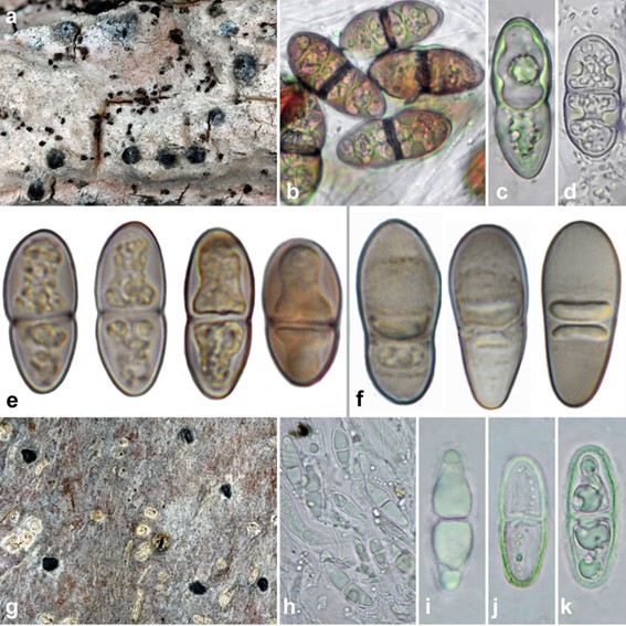
Notes: This new genus is introduced for a single species with highly unusual ascospores that in their mature stage are reminiscent of Arthoniaceae and Graphidaceae rather than Trypetheliaceae. The species was originally described in the genus Distothelia (Schumm and Aptroot 2013), but the ascospores differ clearly in shape and endospore development, resulting in two strongly thickened terminal walls. Immature ascospores appear 3-septate with three eusepta, somewhat similar to ascospores in Novomicrothelia and Pseudobogoriella except for the more numerous septa. However, the two terminal lumina eventually become filled entirely with endospore material and simultaneously inflated, so that the two-remaining central lumina appear much smaller than the thickened terminal walls (Fig. 79f). Given the current distribution of ascospore types among basally diverging lineages in the family, we predict that this taxon represents its own lineage, and placement in Bogoriella, together with the other two species previously placed in Distothelia, would be misleading.

Fig. 79 a–d Bogoriella complexoluminata (holotype). a Thallus with ascomata. b–d Ascospores in various stages of development. e B. isthmospora (Aptroot s.n.), ascospores in various stages of develop- ment. f Schummia angulata (Aptroot 14061), ascospores in various stages of development. g–k Macroconstrictolumina megalateralis (holotype). g Thallus with ascomata. h Hamathecium and ascospores. i–k Ascospores in various stages of development. Scale bars: a, g = 1 mm, b–f, h–k = 10 µm. Photographs by André Aptroot except e and f, by Felix Schumm
Species
